 Movie
Movie Controller


+ Open data

- Basic information
| Entry | Database: PDB / ID: 1qbj | ||||||
|---|---|---|---|---|---|---|---|
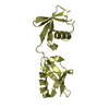
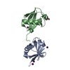
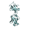
| Title | CRYSTAL STRUCTURE OF THE ZALPHA Z-DNA COMPLEX | ||||||
 Components Components |
| ||||||
 Keywords Keywords | HYDROLASE/DNA / PROTEIN-Z-DNA COMPLEX / HYDROLASE-DNA COMPLEX | ||||||
| Function / homology |  Function and homology information Function and homology informationsomatic diversification of immune receptors via somatic mutation / negative regulation of post-transcriptional gene silencing by regulatory ncRNA / C6 deamination of adenosine / Formation of editosomes by ADAR proteins / double-stranded RNA adenine deaminase / tRNA-specific adenosine deaminase activity / supraspliceosomal complex / double-stranded RNA adenosine deaminase activity / negative regulation of protein kinase activity by regulation of protein phosphorylation / base conversion or substitution editing ...somatic diversification of immune receptors via somatic mutation / negative regulation of post-transcriptional gene silencing by regulatory ncRNA / C6 deamination of adenosine / Formation of editosomes by ADAR proteins / double-stranded RNA adenine deaminase / tRNA-specific adenosine deaminase activity / supraspliceosomal complex / double-stranded RNA adenosine deaminase activity / negative regulation of protein kinase activity by regulation of protein phosphorylation / base conversion or substitution editing / hematopoietic stem cell homeostasis / response to interferon-alpha / adenosine to inosine editing / RISC complex assembly / pre-miRNA processing / negative regulation of hepatocyte apoptotic process / definitive hemopoiesis / negative regulation of type I interferon-mediated signaling pathway / hepatocyte apoptotic process / positive regulation of viral genome replication /  RNA processing / hematopoietic progenitor cell differentiation / protein export from nucleus / erythrocyte differentiation / response to virus / PKR-mediated signaling / mRNA processing / cellular response to virus / osteoblast differentiation / protein import into nucleus / double-stranded RNA binding / Interferon alpha/beta signaling / defense response to virus / innate immune response / nucleolus / DNA binding / RNA binding / nucleoplasm / membrane / metal ion binding / nucleus / cytosol / cytoplasm RNA processing / hematopoietic progenitor cell differentiation / protein export from nucleus / erythrocyte differentiation / response to virus / PKR-mediated signaling / mRNA processing / cellular response to virus / osteoblast differentiation / protein import into nucleus / double-stranded RNA binding / Interferon alpha/beta signaling / defense response to virus / innate immune response / nucleolus / DNA binding / RNA binding / nucleoplasm / membrane / metal ion binding / nucleus / cytosol / cytoplasmSimilarity search - Function | ||||||
| Biological species |  Homo sapiens (human) Homo sapiens (human) | ||||||
| Method | X-RAY DIFFRACTION / SIRAS / Resolution: 2.1 Å | ||||||
 Authors Authors | Schwartz, T. / Rould, M.A. / Rich, A. | ||||||
 Citation Citation | Journal: Science / Year: 1999 Title: Crystal structure of the Zalpha domain of the human editing enzyme ADAR1 bound to left-handed Z-DNA. Authors: Schwartz, T. / Rould, M.A. / Lowenhaupt, K. / Herbert, A. / Rich, A. | ||||||
| History |
|
- Structure visualization
| Structure viewer | Molecule: MolmilJmol/JSmol |
|---|
- Downloads & links
-Download
| PDBx/mmCIF format | 1qbj.cif.gz | 67.5 KB | Display | PDBx/mmCIF format |
|---|---|---|---|---|
| PDB format | pdb1qbj.ent.gz | 47.6 KB | Display | PDB format |
| PDBx/mmJSON format | 1qbj.json.gz | Tree view | PDBx/mmJSON format | |
| Others |  Other downloads Other downloads |
-Validation report
| Arichive directory | https://data.pdbj.org/pub/pdb/validation_reports/qb/1qbjftp://data.pdbj.org/pub/pdb/validation_reports/qb/1qbj | HTTPS FTP |
|---|
-Related structure data
| Similar structure data |
|---|


-Links
 PDBj
PDBj
- Assembly
| Deposited unit | 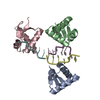
| ||||||||||||||||||||||||||||
|---|---|---|---|---|---|---|---|---|---|---|---|---|---|---|---|---|---|---|---|---|---|---|---|---|---|---|---|---|---|
| 1 | 
| ||||||||||||||||||||||||||||
| 2 | 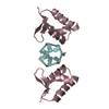
| ||||||||||||||||||||||||||||
| Unit cell |
| ||||||||||||||||||||||||||||
| Components on special symmetry positions |
| ||||||||||||||||||||||||||||
| Noncrystallographic symmetry (NCS) | NCS oper:
|
-Components
| #1: DNA chain | Mass: 2114.398 Da / Num. of mol.: 3 / Source method: obtained synthetically / Details: SYNTHETIC #2: Protein | Mass: 9002.293 Da / Num. of mol.: 3 / Fragment: N-TERMINAL HELIX-TURN-HELIX DOMAIN ZALPHA Source method: isolated from a genetically manipulated source Source: (gene. exp.) Homo sapiens (human) / Plasmid: PET28A / Production host:  Escherichia coli (E. coli) / Strain (production host): NOVABLUE (DE3) / References: UniProt: P55265 Escherichia coli (E. coli) / Strain (production host): NOVABLUE (DE3) / References: UniProt: P55265#3: Water | ChemComp-HOH / | Water Mass: 18.015 Da / Num. of mol.: 244 / Source method: isolated from a natural source / Formula: H2O Mass: 18.015 Da / Num. of mol.: 244 / Source method: isolated from a natural source / Formula: H2O |
|---|
-Experimental details
-Experiment
| Experiment | Method: X-RAY DIFFRACTION / Number of used crystals: 1 |
|---|
- Sample preparation
| Crystal | Density Matthews: 1.97 Å3/Da / Density % sol: 37 % | |||||||||||||||
|---|---|---|---|---|---|---|---|---|---|---|---|---|---|---|---|---|
| Crystal grow | pH: 5.6 Details: HANGING DROP VAPOR DIFFUSION OVER 1.6 M (NH4)2SO4, 10 % GLYCEROL AT 24 DEGREES CELSIUS, pH 5.6 | |||||||||||||||
| Crystal grow | *PLUS Method: vapor diffusion, hanging drop | |||||||||||||||
| Components of the solutions | *PLUS
|
-Data collection
| Diffraction | Mean temperature: 123 K |
|---|---|
| Diffraction source | Source: ROTATING ANODE / Type: RIGAKU RU200 / Wavelength: 1.5418 |
| Detector | Type: RIGAKU RAXIS IIC / Detector: IMAGE PLATE / Date: Apr 20, 1998 |
| Radiation | Protocol: SINGLE WAVELENGTH / Monochromatic (M) / Laue (L): M / Scattering type: x-ray |
| Radiation wavelength | Wavelength: 1.5418 Å / Relative weight: 1 |
| Reflection | Resolution: 2.1→20 Å / Num. obs: 29702 / % possible obs: 99.9 % / Observed criterion σ(I): 0 / Redundancy: 9.8 % / Rsym value: 0.074 / Net I/σ(I): 27.3 |
| Reflection shell | Resolution: 2.1→2.18 Å / Redundancy: 2.9 % / Mean I/σ(I) obs: 2 / Rsym value: 0.535 / % possible all: 99.3 |
| Reflection | *PLUS Rmerge(I) obs: 0.074 |
| Reflection shell | *PLUS % possible obs: 99.3 % / Rmerge(I) obs: 0.535 |
- Processing
| Software |
| ||||||||||||||||||||||||||||||||||||||||||||||||||||||||||||
|---|---|---|---|---|---|---|---|---|---|---|---|---|---|---|---|---|---|---|---|---|---|---|---|---|---|---|---|---|---|---|---|---|---|---|---|---|---|---|---|---|---|---|---|---|---|---|---|---|---|---|---|---|---|---|---|---|---|---|---|---|---|
| Refinement | Method to determine structure: SIRAS / Resolution: 2.1→6 Å / Data cutoff high absF: 100000 / Data cutoff low absF: 0.001 / Cross valid method: THROUGHOUT / σ(F): 0
| ||||||||||||||||||||||||||||||||||||||||||||||||||||||||||||
| Displacement parameters | Biso mean: 29.05 Å2 | ||||||||||||||||||||||||||||||||||||||||||||||||||||||||||||
| Refinement step | Cycle: LAST / Resolution: 2.1→6 Å
| ||||||||||||||||||||||||||||||||||||||||||||||||||||||||||||
| Refine LS restraints |
| ||||||||||||||||||||||||||||||||||||||||||||||||||||||||||||
| LS refinement shell | Resolution: 2.1→2.19 Å / Total num. of bins used: 8
| ||||||||||||||||||||||||||||||||||||||||||||||||||||||||||||
| Xplor file |
| ||||||||||||||||||||||||||||||||||||||||||||||||||||||||||||
| Software | *PLUS Name: X-PLOR / Version: 3.851 / Classification: refinement | ||||||||||||||||||||||||||||||||||||||||||||||||||||||||||||
| Refine LS restraints | *PLUS
|
