[English] 日本語
Yorodumi
- PDB-1w7t: Photoproduct of the Wild-Type Aequorea victoria Green Fluorescent... -

+
Open data


ID or keywords:

Loading...

-
Basic information

Entry
Database: PDB / ID: 1w7t
TitlePhotoproduct of the Wild-Type Aequorea victoria Green Fluorescent Protein at 100 K
ComponentsGREEN FLUORESCENT PROTEIN
KeywordsLUMINESCENT PROTEIN / BETA-BARREL / BIOLUMINESCENCE / PHOTOPRODUCT
Function / homology
Function and homology information


bioluminescence / generation of precursor metabolites and energy
Similarity search - Function
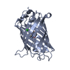
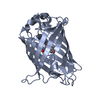
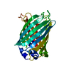
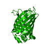
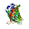
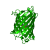
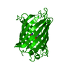
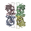
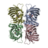
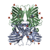
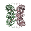
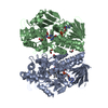
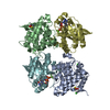
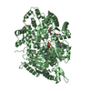
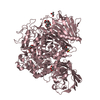
Green Fluorescent Protein / Green fluorescent protein / Green fluorescent protein, GFP / Green fluorescent protein-related / Green fluorescent protein / Green fluorescent protein / Beta Barrel / Mainly Beta
Similarity search - Domain/homology
Green fluorescent protein
Similarity search - Component
Biological speciesAEQUOREA VICTORIA (jellyfish)
MethodX-RAY DIFFRACTION / SYNCHROTRON / MOLECULAR REPLACEMENT / Resolution: 1.85 Å
AuthorsVan Thor, J.J. / Georgiev, G.Y. / Towrie, M. / Sage, J.T.
CitationJournal: J.Biol.Chem. / Year: 2005
Title: Ultrafast and Low Barrier Motions in the Photoreactions of the Green Fluorescent Protein
Authors: Van Thor, J.J. / Georgiev, G.Y. / Towrie, M. / Sage, J.T.
History
DepositionSep 9, 2004Deposition site: PDBE / Processing site: PDBE
Revision 1.0Jul 20, 2005Provider: repository / Type: Initial release
Revision 1.1Mar 5, 2014Group: Atomic model / Database references ...Atomic model / Database references / Derived calculations / Non-polymer description / Other / Source and taxonomy / Structure summary / Version format compliance
Revision 1.2May 15, 2019Group: Data collection / Derived calculations ...Data collection / Derived calculations / Experimental preparation / Other
Category: exptl_crystal_grow / pdbx_database_proc ...exptl_crystal_grow / pdbx_database_proc / pdbx_database_status / struct_conn
Item: _exptl_crystal_grow.temp / _pdbx_database_status.recvd_author_approval / _struct_conn.pdbx_leaving_atom_flag
Revision 1.3Oct 23, 2019Group: Data collection / Database references / Other / Category: pdbx_database_status / struct_ref_seq_dif
Item: _pdbx_database_status.status_code_sf / _struct_ref_seq_dif.details
Revision 2.0Nov 15, 2023Group: Atomic model / Data collection ...Atomic model / Data collection / Database references / Derived calculations
Category: atom_site / chem_comp_atom ...atom_site / chem_comp_atom / chem_comp_bond / database_2 / pdbx_validate_main_chain_plane / pdbx_validate_rmsd_angle / pdbx_validate_rmsd_bond / struct_conn
Item: _atom_site.auth_atom_id / _atom_site.label_atom_id ..._atom_site.auth_atom_id / _atom_site.label_atom_id / _database_2.pdbx_DOI / _database_2.pdbx_database_accession / _struct_conn.pdbx_leaving_atom_flag / _struct_conn.ptnr1_label_atom_id / _struct_conn.ptnr2_label_atom_id
Revision 2.1Dec 13, 2023Group: Refinement description / Category: pdbx_initial_refinement_model
Remark 700 
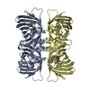
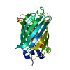
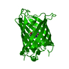
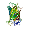
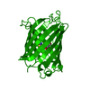
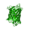
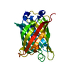
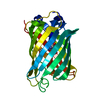
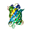
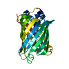
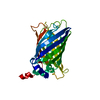
SHEET DETERMINATION METHOD: DSSP THE SHEETS PRESENTED AS "AA, BA" IN EACH CHAIN ON SHEET RECORDS ... SHEET DETERMINATION METHOD: DSSP THE SHEETS PRESENTED AS "AA, BA" IN EACH CHAIN ON SHEET RECORDS BELOW IS ACTUALLY AN 12-STRANDED BARREL THIS IS REPRESENTED BY A 13-STRANDED SHEET IN WHICH THE FIRST AND LAST STRANDS ARE IDENTICAL. THE SHEETS PRESENTED AS "CA, DA" IN EACH CHAIN ON SHEET RECORDS BELOW IS ACTUALLY AN 10-STRANDED BARREL THIS IS REPRESENTED BY A 11-STRANDED SHEET IN WHICH THE FIRST AND LAST STRANDS ARE IDENTICAL.

-
Structure visualization

Structure viewerMolecule:
MolmilJmol/JSmol

Downloads & links

-
Assembly

Deposited unit
A: GREEN FLUORESCENT PROTEIN
B: GREEN FLUORESCENT PROTEIN
C: GREEN FLUORESCENT PROTEIN
D: GREEN FLUORESCENT PROTEIN


Theoretical massNumber of molelcules
Total (without water)107,5494
Polymers107,5494
Non-polymers00
Water16,376909
1


  • Idetical with deposited unit
  • defined by author&software
TypeNameSymmetry operationNumber
identity operation1_555x,y,z1
Buried area6880 Å2
ΔGint-41.4 kcal/mol
Surface area44960 Å2
MethodPQS
Unit cell
Length a, b, c (Å)155.308, 52.815, 141.944
Angle α, β, γ (deg.)90.00, 120.07, 90.00
Int Tables number5
Space group name H-MC121
Components on special symmetry positions
IDModelComponents
11A-2195-

HOH

-
Components

#1: Protein
GREEN FLUORESCENT PROTEIN /


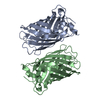
Mass: 26887.346 Da / Num. of mol.: 4 / Mutation: YES
Source method: isolated from a genetically manipulated source
Details: THE CHROMOPHORE, GYS, IS PART OF THE PEPTIDE CHAIN BETWEEN RESIDUES 64 AND 68.
Source: (gene. exp.) AEQUOREA VICTORIA (jellyfish) / Tissue: CIRCUMORAL RING CANAL / Organ: PHOTOGENIC ORGAN / Production host: ESCHERICHIA COLI M15 (bacteria) / References: UniProt: P42212
#2: Water ChemComp-HOH / water / Water


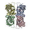
Mass: 18.015 Da / Num. of mol.: 909 / Source method: isolated from a natural source / Formula: H2O
Compound detailsENGINEERED MUTATION IN CHAINS A, B, C, D, GLN 80 TO ARG
Sequence detailsMODRES: 1W7T GLU 222 SIDECHAIN IS SPECIFICALLY DECARBOXYLATED AS A RESULT OF PHOTOTRANSFORMATION. ...MODRES: 1W7T GLU 222 SIDECHAIN IS SPECIFICALLY DECARBOXYLATED AS A RESULT OF PHOTOTRANSFORMATION. OE1, OE2, CD ATOMS ARE NOT PRESENT FOR THIS RESIDUE IN ALL CHAINS

-
Experimental details

-
Experiment

ExperimentMethod: X-RAY DIFFRACTION / Number of used crystals: 1

-
Sample preparation

CrystalDensity Matthews: 2.2 Å3/Da / Density % sol: 43 %
Crystal growTemperature: 277 K / pH: 7.8
Details: CRYSTALS WERE GROWN AT 4C FROM 50 MM MGCL2, 14-17 % PEG3350 AND 50-100 MM TRIS/CL PH 7.8 - 8.6.

-
Data collection

DiffractionMean temperature: 100 K
Diffraction sourceSource: SYNCHROTRON / Site: SRS / Beamline: PX14.2 / Wavelength: 0.97809
DetectorType: ADSC CCD / Detector: CCD / Date: Jul 8, 2003 / Details: RH COATED SILICON MIRROR
RadiationMonochromator: SILICON (111) / Protocol: SINGLE WAVELENGTH / Monochromatic (M) / Laue (L): M / Scattering type: x-ray
Radiation wavelengthWavelength: 0.97809 Å / Relative weight: 1
ReflectionResolution: 1.85→36.76 Å / Num. obs: 85530 / % possible obs: 97.6 % / Observed criterion σ(I): 0 / Redundancy: 3.7 % / Rmerge(I) obs: 0.084 / Net I/σ(I): 5.5
Reflection shellResolution: 1.85→1.95 Å / Redundancy: 2.7 % / Rmerge(I) obs: 0.248 / Mean I/σ(I) obs: 2.9 / % possible all: 86.6

-
Processing

Software
NameVersionClassification
REFMAC5.1.24refinement
MOSFLMdata reduction
SCALAdata scaling
MOLREPphasing
RefinementMethod to determine structure: MOLECULAR REPLACEMENT
Starting model: PDB ENTRY 1HCJ
Resolution: 1.85→119.52 Å / Cor.coef. Fo:Fc: 0.944 / Cor.coef. Fo:Fc free: 0.928 / SU B: 2.758 / SU ML: 0.084 / Cross valid method: THROUGHOUT / ESU R: 0.144 / ESU R Free: 0.134 / Stereochemistry target values: MAXIMUM LIKELIHOOD / Details: HYDROGENS HAVE BEEN ADDED IN THE RIDING POSITIONS
RfactorNum. reflection% reflectionSelection details
Rfree0.22 4162 5 %RANDOM
Rwork0.18 ---
obs0.182 79139 97.4 %-
Solvent computationIon probe radii: 0.8 Å / Shrinkage radii: 0.8 Å / VDW probe radii: 1.4 Å / Solvent model: BABINET MODEL WITH MASK
Displacement parametersBiso mean: 16.25 Å2
Baniso -1Baniso -2Baniso -3
1--0.21 Å20 Å2-0.26 Å2
2--0.62 Å20 Å2
3----0.68 Å2
Refinement stepCycle: LAST / Resolution: 1.85→119.52 Å
ProteinNucleic acidLigandSolventTotal
Num. atoms7302 0 0 909 8211
Refine LS restraints
Refine-IDTypeDev idealDev ideal targetNumber
X-RAY DIFFRACTIONr_bond_refined_d0.0130.0217488
X-RAY DIFFRACTIONr_bond_other_d0.0030.026578
X-RAY DIFFRACTIONr_angle_refined_deg1.5091.95610119
X-RAY DIFFRACTIONr_angle_other_deg1.223315456
X-RAY DIFFRACTIONr_dihedral_angle_1_deg6.5695899
X-RAY DIFFRACTIONr_dihedral_angle_2_deg
X-RAY DIFFRACTIONr_dihedral_angle_3_deg
X-RAY DIFFRACTIONr_dihedral_angle_4_deg
X-RAY DIFFRACTIONr_chiral_restr0.1260.21108
X-RAY DIFFRACTIONr_gen_planes_refined0.0080.028277
X-RAY DIFFRACTIONr_gen_planes_other0.0070.021475
X-RAY DIFFRACTIONr_nbd_refined0.2510.21242
X-RAY DIFFRACTIONr_nbd_other0.2620.27846
X-RAY DIFFRACTIONr_nbtor_refined
X-RAY DIFFRACTIONr_nbtor_other0.0840.24489
X-RAY DIFFRACTIONr_xyhbond_nbd_refined0.1440.2625
X-RAY DIFFRACTIONr_xyhbond_nbd_other
X-RAY DIFFRACTIONr_metal_ion_refined
X-RAY DIFFRACTIONr_metal_ion_other
X-RAY DIFFRACTIONr_symmetry_vdw_refined0.1250.210
X-RAY DIFFRACTIONr_symmetry_vdw_other0.2480.214
X-RAY DIFFRACTIONr_symmetry_hbond_refined0.1130.215
X-RAY DIFFRACTIONr_symmetry_hbond_other
X-RAY DIFFRACTIONr_symmetry_metal_ion_refined
X-RAY DIFFRACTIONr_symmetry_metal_ion_other
X-RAY DIFFRACTIONr_mcbond_it0.7741.54492
X-RAY DIFFRACTIONr_mcbond_other
X-RAY DIFFRACTIONr_mcangle_it1.53527274
X-RAY DIFFRACTIONr_mcangle_other
X-RAY DIFFRACTIONr_scbond_it2.60532996
X-RAY DIFFRACTIONr_scbond_other
X-RAY DIFFRACTIONr_scangle_it4.3514.52844
X-RAY DIFFRACTIONr_scangle_other
X-RAY DIFFRACTIONr_long_range_B_refined
X-RAY DIFFRACTIONr_long_range_B_other
X-RAY DIFFRACTIONr_rigid_bond_restr
X-RAY DIFFRACTIONr_sphericity_free
X-RAY DIFFRACTIONr_sphericity_bonded
LS refinement shellResolution: 1.85→1.9 Å / Total num. of bins used: 20 /
RfactorNum. reflection
Rfree0.266 244
Rwork0.201 4817

+
About Yorodumi

-
News

-
Feb 9, 2022. New format data for meta-information of EMDB entries

New format data for meta-information of EMDB entries

  • Version 3 of the EMDB header file is now the official format.
  • The previous official version 1.9 will be removed from the archive.

Related info.:EMDB header

External links:wwPDB to switch to version 3 of the EMDB data model

-
Aug 12, 2020. Covid-19 info

Covid-19 info

URL: https://pdbj.org/emnavi/covid19.php

New page: Covid-19 featured information page in EM Navigator.

Related info.:Covid-19 info / Mar 5, 2020. Novel coronavirus structure data

+
Mar 5, 2020. Novel coronavirus structure data

Novel coronavirus structure data

Related info.:Yorodumi Speices / Aug 12, 2020. Covid-19 info

External links:COVID-19 featured content - PDBj / Molecule of the Month (242):Coronavirus Proteases

+
Jan 31, 2019. EMDB accession codes are about to change! (news from PDBe EMDB page)

EMDB accession codes are about to change! (news from PDBe EMDB page)

  • The allocation of 4 digits for EMDB accession codes will soon come to an end. Whilst these codes will remain in use, new EMDB accession codes will include an additional digit and will expand incrementally as the available range of codes is exhausted. The current 4-digit format prefixed with “EMD-” (i.e. EMD-XXXX) will advance to a 5-digit format (i.e. EMD-XXXXX), and so on. It is currently estimated that the 4-digit codes will be depleted around Spring 2019, at which point the 5-digit format will come into force.
  • The EM Navigator/Yorodumi systems omit the EMD- prefix.

Related info.:Q: What is EMD? / ID/Accession-code notation in Yorodumi/EM Navigator

External links:EMDB Accession Codes are Changing Soon! / Contact to PDBj

+
Jul 12, 2017. Major update of PDB

Major update of PDB

  • wwPDB released updated PDB data conforming to the new PDBx/mmCIF dictionary.
  • This is a major update changing the version number from 4 to 5, and with Remediation, in which all the entries are updated.
  • In this update, many items about electron microscopy experimental information are reorganized (e.g. em_software).
  • Now, EM Navigator and Yorodumi are based on the updated data.

External links:wwPDB Remediation / Enriched Model Files Conforming to OneDep Data Standards Now Available in the PDB FTP Archive

-
Yorodumi

Thousand views of thousand structures

  • Yorodumi is a browser for structure data from EMDB, PDB, SASBDB, etc.
  • This page is also the successor to EM Navigator detail page, and also detail information page/front-end page for Omokage search.
  • The word "yorodu" (or yorozu) is an old Japanese word meaning "ten thousand". "mi" (miru) is to see.

Related info.:EMDB / PDB / SASBDB / Comparison of 3 databanks / Yorodumi Search / Aug 31, 2016. New EM Navigator & Yorodumi / Yorodumi Papers / Jmol/JSmol / Function and homology information / Changes in new EM Navigator and Yorodumi

Read more