 Movie
Movie Controller


+ Open data

- Basic information
| Entry | Database: PDB / ID: 1emc | |||||||||
|---|---|---|---|---|---|---|---|---|---|---|
| Title | GREEN FLUORESCENT PROTEIN FROM AEQUOREA VICTORIA, MUTANT | |||||||||
 Components Components | GREEN FLUORESCENT PROTEIN | |||||||||
 Keywords Keywords | LUMINESCENCE / FLUORESCENT PROTEIN / BETA-BARREL / BIOLUMINESCENCE | |||||||||
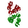
| Function / homology |  Function and homology information Function and homology information | |||||||||
| Biological species |   Aequorea victoria (jellyfish) Aequorea victoria (jellyfish) | |||||||||
| Method | X-RAY DIFFRACTION / MOLECULAR REPLACEMENT / Resolution: 2.3 Å | |||||||||
 Authors Authors | Palm, G. / Zdanov, A. / Wlodawer, A. | |||||||||
 Citation Citation | Journal: Nat.Struct.Biol. / Year: 1997 Title: The structural basis for spectral variations in green fluorescent protein. Authors: Palm, G.J. / Zdanov, A. / Gaitanaris, G.A. / Stauber, R. / Pavlakis, G.N. / Wlodawer, A. | |||||||||
| History |
|
- Structure visualization
| Structure viewer | Molecule: MolmilJmol/JSmol |
|---|
- Downloads & links
-Download
| PDBx/mmCIF format | 1emc.cif.gz | 188.5 KB | Display | PDBx/mmCIF format |
|---|---|---|---|---|
| PDB format | pdb1emc.ent.gz | 150.5 KB | Display | PDB format |
| PDBx/mmJSON format | 1emc.json.gz | Tree view | PDBx/mmJSON format | |
| Others |  Other downloads Other downloads |
-Validation report
| Arichive directory | https://data.pdbj.org/pub/pdb/validation_reports/em/1emcftp://data.pdbj.org/pub/pdb/validation_reports/em/1emc | HTTPS FTP |
|---|
-Related structure data
| Related structure data | 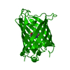 1emeC  1emfC 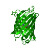 1emkC 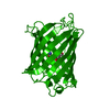 1emlC  1emmC  2emdC 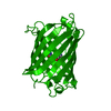 2emnC 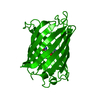 2emoC  1emaS S: Starting model for refinement C: citing same article ( |
|---|---|
| Similar structure data |
-Links
 PDBj
PDBj

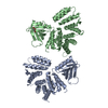
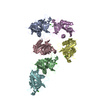
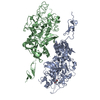
- Assembly
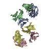
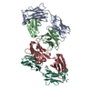
| Deposited unit | 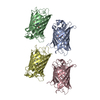
| |||||||||||||||||||||||||||||||
|---|---|---|---|---|---|---|---|---|---|---|---|---|---|---|---|---|---|---|---|---|---|---|---|---|---|---|---|---|---|---|---|---|
| 1 |
| |||||||||||||||||||||||||||||||
| Unit cell |
| |||||||||||||||||||||||||||||||
| Noncrystallographic symmetry (NCS) | NCS domain:
NCS oper:
|
-Components
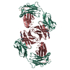
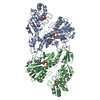
| #1: Protein | / GFP Mass: 26941.283 Da / Num. of mol.: 4 / Mutation: INS(A1[B]), F64L, I167T, K238N Source method: isolated from a genetically manipulated source Source: (gene. exp.) Aequorea victoria (jellyfish) / Tissue: CIRCUMORAL RING CANAL / Gene: GFP / Organ: PHOTOGENIC ORGAN / Plasmid: PFRED11 / Species (production host): Escherichia coli / Cellular location (production host): CYTOPLASM / Gene (production host): SG11 / Production host:  Escherichia coli BL21 (bacteria) / Strain (production host): BL21 / References: UniProt: P42212 Escherichia coli BL21 (bacteria) / Strain (production host): BL21 / References: UniProt: P42212#2: Water | ChemComp-HOH / | Water Mass: 18.015 Da / Num. of mol.: 158 / Source method: isolated from a natural source / Formula: H2O Mass: 18.015 Da / Num. of mol.: 158 / Source method: isolated from a natural source / Formula: H2ONonpolymer details | GYS: [(4Z)-2-[(1R)-1-AMINO-2-HYDROXYETHYL]-4-(4-HYDROXYBENZYLIDENE)-5-OXO-4,5-DIHYDRO-1H-IMIDAZOL-1- ...GYS: [(4Z)-2-[(1R)-1-AMINO-2-HYDROXYETH | |
|---|
-Experimental details
-Experiment
| Experiment | Method: X-RAY DIFFRACTION / Number of used crystals: 1 |
|---|
- Sample preparation
| Crystal | Density Matthews: 2.25 Å3/Da / Density % sol: 47 % | ||||||||||||||||||||||||||||||
|---|---|---|---|---|---|---|---|---|---|---|---|---|---|---|---|---|---|---|---|---|---|---|---|---|---|---|---|---|---|---|---|
| Crystal grow | Method: vapor diffusion, hanging drop / pH: 8 Details: PROTEIN WAS CRYSTALLIZED BY HANGING DROP METHOD. PROTEIN SOLUTION: 14 MG/ML IN 20 MM KPO4, PH 7.0 WELL SOLUTION: 60% MPD, 50MM TRISCL, PH 8.0 PROTEIN:WELL 1:1, vapor diffusion - hanging drop PH range: 7.0-8.0 | ||||||||||||||||||||||||||||||
| Crystal grow | *PLUS Method: vapor diffusion, hanging dropDetails: protein solution is mixed in a 1:1 ratio with well solution | ||||||||||||||||||||||||||||||
| Components of the solutions | *PLUS
|
-Data collection
| Diffraction | Mean temperature: 295 K |
|---|---|
| Diffraction source | Source: ROTATING ANODE / Type: RIGAKU / Wavelength: 1.5418 |
| Detector | Type: MACSCIENCE / Detector: IMAGE PLATE / Date: Oct 12, 1995 / Details: MIRROR |
| Radiation | Monochromator: NI / Monochromatic (M) / Laue (L): M / Scattering type: x-ray |
| Radiation wavelength | Wavelength: 1.5418 Å / Relative weight: 1 |
| Reflection | Resolution: 2.3→10 Å / Num. obs: 31013 / % possible obs: 78.5 % / Observed criterion σ(I): 0 / Redundancy: 2.9 % / Rsym value: 0.078 / Net I/σ(I): 6.5 |
| Reflection shell | Resolution: 2.3→2.38 Å / Mean I/σ(I) obs: 1.9 / Rsym value: 0.253 / % possible all: 56.9 |
| Reflection | *PLUS Rmerge(I) obs: 0.078 |
| Reflection shell | *PLUS % possible obs: 56.9 % / Rmerge(I) obs: 0.253 |
- Processing
| Software |
| ||||||||||||||||||||||||||||||||||||||||||||||||||||||||||||||||||||||||||||||||
|---|---|---|---|---|---|---|---|---|---|---|---|---|---|---|---|---|---|---|---|---|---|---|---|---|---|---|---|---|---|---|---|---|---|---|---|---|---|---|---|---|---|---|---|---|---|---|---|---|---|---|---|---|---|---|---|---|---|---|---|---|---|---|---|---|---|---|---|---|---|---|---|---|---|---|---|---|---|---|---|---|---|
| Refinement | Method to determine structure: MOLECULAR REPLACEMENT Starting model: PDB ENTRY 1EMA Resolution: 2.3→10 Å / Rfactor Rfree error: 0.006 / Data cutoff high absF: 100000 / Data cutoff low absF: 0.1 / Isotropic thermal model: RESTRAINED / σ(F): 2 Details: PARAMETERS FOR THE CHROMOPHORE WERE ESTIMATED ACCORDING TO A MODEL COMPOUND (B.TINANT ET AL., CRYST. STRUCT. COMM., 1980, 9, 671-674). ATOMS CA, CB, OG1, AND CG2 OF THR ? 203 ? WERE REFINED ...Details: PARAMETERS FOR THE CHROMOPHORE WERE ESTIMATED ACCORDING TO A MODEL COMPOUND (B.TINANT ET AL., CRYST. STRUCT. COMM., 1980, 9, 671-674). ATOMS CA, CB, OG1, AND CG2 OF THR ? 203 ? WERE REFINED IN TWO ALTERNATE CONFORMATIONS.
| ||||||||||||||||||||||||||||||||||||||||||||||||||||||||||||||||||||||||||||||||
| Displacement parameters | Biso mean: 15.4 Å2 | ||||||||||||||||||||||||||||||||||||||||||||||||||||||||||||||||||||||||||||||||
| Refinement step | Cycle: LAST / Resolution: 2.3→10 Å
| ||||||||||||||||||||||||||||||||||||||||||||||||||||||||||||||||||||||||||||||||
| Refine LS restraints |
| ||||||||||||||||||||||||||||||||||||||||||||||||||||||||||||||||||||||||||||||||
| Refine LS restraints NCS |
| ||||||||||||||||||||||||||||||||||||||||||||||||||||||||||||||||||||||||||||||||
| LS refinement shell | Resolution: 2.3→2.4 Å / Rfactor Rfree error: 0.026 / Total num. of bins used: 8
| ||||||||||||||||||||||||||||||||||||||||||||||||||||||||||||||||||||||||||||||||
| Xplor file |
| ||||||||||||||||||||||||||||||||||||||||||||||||||||||||||||||||||||||||||||||||
| Software | *PLUS Name: X-PLOR / Version: 3.1 / Classification: refinement | ||||||||||||||||||||||||||||||||||||||||||||||||||||||||||||||||||||||||||||||||
| Refinement | *PLUS | ||||||||||||||||||||||||||||||||||||||||||||||||||||||||||||||||||||||||||||||||
| Solvent computation | *PLUS | ||||||||||||||||||||||||||||||||||||||||||||||||||||||||||||||||||||||||||||||||
| Displacement parameters | *PLUS | ||||||||||||||||||||||||||||||||||||||||||||||||||||||||||||||||||||||||||||||||
| Refine LS restraints | *PLUS
| ||||||||||||||||||||||||||||||||||||||||||||||||||||||||||||||||||||||||||||||||
| LS refinement shell | *PLUS Rfactor obs: 0.27 |
