 Movie
Movie Controller


+ Open data

- Basic information
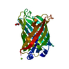
| Entry | Database: PDB / ID: 1yfp | ||||||
|---|---|---|---|---|---|---|---|
| Title | STRUCTURE OF YELLOW-EMISSION VARIANT OF GFP | ||||||
 Components Components | YELLOW FLUORESCENT PROTEIN | ||||||
 Keywords Keywords | LUMINESCENCE / GREEN FLUORESCENT PROTEIN / YELLOW-EMISSION VARIANT / BIOLUMINESCENCE / PHOTOACTIVE PROTEIN / FLUORESCENT TAG | ||||||
| Function / homology |  Function and homology information Function and homology information | ||||||
| Biological species |   Aequorea victoria (jellyfish) Aequorea victoria (jellyfish) | ||||||
| Method | X-RAY DIFFRACTION / MOLECULAR REPLACEMENT / Resolution: 2.5 Å | ||||||
 Authors Authors | Wachter, R.M. / Elsliger, M.-A. / Kallio, K. / Hanson, G.T. / Remington, S.J. | ||||||
 Citation Citation | Journal: Structure / Year: 1998 Title: Structural basis of spectral shifts in the yellow-emission variants of green fluorescent protein. Authors: Wachter, R.M. / Elsliger, M.A. / Kallio, K. / Hanson, G.T. / Remington, S.J. | ||||||
| History |
| ||||||
| Remark 999 | THE CHROMOPHORE CRO IS GENERATED BY AN AUTOCATALYTIC CYCLIZATION OF THE POLYPEPTIDE BACKBONE OF SER ...THE CHROMOPHORE CRO IS GENERATED BY AN AUTOCATALYTIC CYCLIZATION OF THE POLYPEPTIDE BACKBONE OF SER 65, TYR 66, GLY 67. RESIDUES SER65 under went mutation to GLY65 before cyclization. |
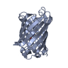
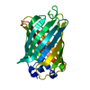
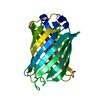
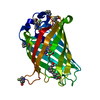
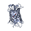
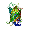
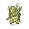
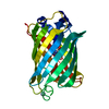
- Structure visualization
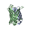
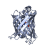
| Structure viewer | Molecule: MolmilJmol/JSmol |
|---|
- Downloads & links
-Download
| PDBx/mmCIF format | 1yfp.cif.gz | 94.5 KB | Display | PDBx/mmCIF format |
|---|---|---|---|---|
| PDB format | pdb1yfp.ent.gz | 75.2 KB | Display | PDB format |
| PDBx/mmJSON format | 1yfp.json.gz | Tree view | PDBx/mmJSON format | |
| Others |  Other downloads Other downloads |
-Validation report
| Arichive directory | https://data.pdbj.org/pub/pdb/validation_reports/yf/1yfpftp://data.pdbj.org/pub/pdb/validation_reports/yf/1yfp | HTTPS FTP |
|---|
-Related structure data
| Related structure data |  2yfpC  1emaS S: Starting model for refinement C: citing same article ( |
|---|---|
| Similar structure data |


-Links
 PDBj
PDBj

- Assembly
| Deposited unit | 
| ||||||||
|---|---|---|---|---|---|---|---|---|---|
| 1 | 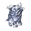
| ||||||||
| 2 | 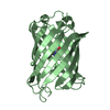
| ||||||||
| Unit cell |
| ||||||||
| Noncrystallographic symmetry (NCS) | NCS oper: (Code: given Matrix: (-0.99998, -0.00309, -0.00619), Vector : |
-Components
| #1: Protein | Mass: 25709.994 Da / Num. of mol.: 2 / Mutation: S65G, V68L, S72A, Q80R, H148G, T203Y Source method: isolated from a genetically manipulated source Source: (gene. exp.) Aequorea victoria (jellyfish) / Tissue: CIRCUMORAL RING CANAL / Gene: GFP / Organ: PHOTOGENIC ORGAN / Plasmid: PRSETB (INVITROGEN) / Cellular location (production host): CYTOPLASM / Production host:  Escherichia coli (E. coli) / Strain (production host): JM109 (DE3) / References: UniProt: P42212 Escherichia coli (E. coli) / Strain (production host): JM109 (DE3) / References: UniProt: P42212#2: Water | ChemComp-HOH / | Water Mass: 18.015 Da / Num. of mol.: 102 / Source method: isolated from a natural source / Formula: H2O Mass: 18.015 Da / Num. of mol.: 102 / Source method: isolated from a natural source / Formula: H2O |
|---|
-Experimental details
-Experiment
| Experiment | Method: X-RAY DIFFRACTION / Number of used crystals: 2 |
|---|
- Sample preparation
| Crystal | Density Matthews: 2.66 Å3/Da / Density % sol: 53.4 % / Description: ISOMORPHOUS REPLACEMENT | ||||||||||||||||||||
|---|---|---|---|---|---|---|---|---|---|---|---|---|---|---|---|---|---|---|---|---|---|
| Crystal grow | Temperature: 288 K / Method: vapor diffusion, hanging drop / pH: 7.5 Details: YFP WAS CONCENTRATED TO 10 MG/ML IN 50 MM HEPES PH 7.5. ROD-SHAPED CRYSTALS WITH APPROXIMATE DIMENSIONS OF 0.8 X 0.12 X 0.03 MM WERE GROWN IN HANGING DROPS CONTAINING 5 MICROLITERS PROTEIN ...Details: YFP WAS CONCENTRATED TO 10 MG/ML IN 50 MM HEPES PH 7.5. ROD-SHAPED CRYSTALS WITH APPROXIMATE DIMENSIONS OF 0.8 X 0.12 X 0.03 MM WERE GROWN IN HANGING DROPS CONTAINING 5 MICROLITERS PROTEIN AND 5 MICROLITERS MOTHER LIQUOR AT 15 DEGREES C AFTER 2 WEEKS. THE MOTHER LIQUOR CONTAINED 2.2 M SODIUM/POTASSIUM PHOSPHATE PH 6.9., vapor diffusion - hanging drop, temperature 288K | ||||||||||||||||||||
| Crystal | *PLUS | ||||||||||||||||||||
| Crystal grow | *PLUS Temperature: 15 ℃ / Method: vapor diffusion, hanging drop | ||||||||||||||||||||
| Components of the solutions | *PLUS
|
-Data collection
| Diffraction | Mean temperature: 295 K |
|---|---|
| Diffraction source | Source: ROTATING ANODE / Type: RIGAKU RUH3R / Wavelength: 1.5418 |
| Detector | Type: RIGAKU / Detector: IMAGE PLATE / Date: Dec 5, 1997 / Details: MIRRORS |
| Radiation | Monochromatic (M) / Laue (L): M / Scattering type: x-ray |
| Radiation wavelength | Wavelength: 1.5418 Å / Relative weight: 1 |
| Reflection | Resolution: 2.5→20 Å / Num. obs: 18916 / % possible obs: 92 % / Observed criterion σ(I): 1.9 / Redundancy: 4 % / Biso Wilson estimate: 28.2 Å2 / Rmerge(I) obs: 0.08 / Net I/σ(I): 10.8 |
| Reflection shell | Resolution: 2.5→2.56 Å / Redundancy: 4 % / % possible all: 94 |
| Reflection | *PLUS Num. measured all: 53039 / Rmerge(I) obs: 0.08 |
| Reflection shell | *PLUS % possible obs: 94 % |
- Processing
| Software |
| ||||||||||||||||||||||||||||||||||||||||||||||||||
|---|---|---|---|---|---|---|---|---|---|---|---|---|---|---|---|---|---|---|---|---|---|---|---|---|---|---|---|---|---|---|---|---|---|---|---|---|---|---|---|---|---|---|---|---|---|---|---|---|---|---|---|
| Refinement | Method to determine structure: MOLECULAR REPLACEMENT Starting model: PDB ENTRY 1EMA Resolution: 2.5→20 Å / Isotropic thermal model: TNT / Stereochemistry target values: TNT
| ||||||||||||||||||||||||||||||||||||||||||||||||||
| Solvent computation | Bsol: 150 Å2 / ksol: 0.82 e/Å3 | ||||||||||||||||||||||||||||||||||||||||||||||||||
| Refinement step | Cycle: LAST / Resolution: 2.5→20 Å
| ||||||||||||||||||||||||||||||||||||||||||||||||||
| Refine LS restraints |
| ||||||||||||||||||||||||||||||||||||||||||||||||||
| Software | *PLUS Name: TNT / Version: 5F / Classification: refinement | ||||||||||||||||||||||||||||||||||||||||||||||||||
| Refinement | *PLUS Rfactor obs: 0.192 | ||||||||||||||||||||||||||||||||||||||||||||||||||
| Solvent computation | *PLUS | ||||||||||||||||||||||||||||||||||||||||||||||||||
| Displacement parameters | *PLUS | ||||||||||||||||||||||||||||||||||||||||||||||||||
| Refine LS restraints | *PLUS
|
