 Movie
Movie Controller


+ Open data

- Basic information
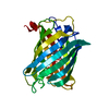
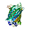
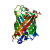
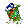
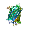
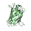
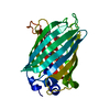
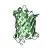
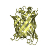
| Entry | Database: PDB / ID: 2yfp | ||||||
|---|---|---|---|---|---|---|---|
| Title | STRUCTURE OF YELLOW-EMISSION VARIANT OF GFP | ||||||
 Components Components | PROTEIN (GREEN FLUORESCENT PROTEIN) | ||||||
 Keywords Keywords |  PHOTOSYNTHESIS / GREEN FLUORESCENT PROTEIN / YELLOW-EMISSION VARIANT / BIOLUMINESCENCE / PHOTOACTIVE PROTEIN / FLUORESCENT TAG PHOTOSYNTHESIS / GREEN FLUORESCENT PROTEIN / YELLOW-EMISSION VARIANT / BIOLUMINESCENCE / PHOTOACTIVE PROTEIN / FLUORESCENT TAG | ||||||
| Function / homology |  Function and homology information Function and homology information | ||||||
| Biological species |   Aequorea victoria (jellyfish) Aequorea victoria (jellyfish) | ||||||
| Method | X-RAY DIFFRACTION / MOLECULAR REPLACEMENT / Resolution: 2.6 Å | ||||||
 Authors Authors | Wachter, R.M. / Elsliger, M.A. / Kallio, K. / Hanson, G.T. / Remington, S.J. | ||||||
 Citation Citation | Journal: Structure / Year: 1998 Title: Structural basis of spectral shifts in the yellow-emission variants of green fluorescent protein. Authors: Wachter, R.M. / Elsliger, M.A. / Kallio, K. / Hanson, G.T. / Remington, S.J. | ||||||
| History |
|
- Structure visualization
| Structure viewer | Molecule: MolmilJmol/JSmol |
|---|
- Downloads & links
-Download
| PDBx/mmCIF format | 2yfp.cif.gz | 58.1 KB | Display | PDBx/mmCIF format |
|---|---|---|---|---|
| PDB format | pdb2yfp.ent.gz | 40.9 KB | Display | PDB format |
| PDBx/mmJSON format | 2yfp.json.gz | Tree view | PDBx/mmJSON format | |
| Others |  Other downloads Other downloads |
-Validation report
| Arichive directory | https://data.pdbj.org/pub/pdb/validation_reports/yf/2yfpftp://data.pdbj.org/pub/pdb/validation_reports/yf/2yfp | HTTPS FTP |
|---|
-Related structure data
| Related structure data |  1yfpC  1emaS S: Starting model for refinement C: citing same article ( |
|---|---|
| Similar structure data |


-Links
 PDBj
PDBj

- Assembly
| Deposited unit | 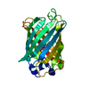
| ||||||||
|---|---|---|---|---|---|---|---|---|---|
| 1 |
| ||||||||
| Unit cell |
|
-Components
| #1: Protein | Mass: 26880.330 Da / Num. of mol.: 1 / Mutation: S65G,V68L,S72A,T203Y,H148G,Q80R Source method: isolated from a genetically manipulated source Source: (gene. exp.) Aequorea victoria (jellyfish) / Tissue: CIRCUMORAL RING CANAL / Gene: GFP / Organ: PHOTOGENIC ORGAN / Plasmid: PRSETB (INVITROGEN) / Cellular location (production host): CYTOPLASM / Production host:  Escherichia coli (E. coli) / Strain (production host): JM109 (DE3) / References: UniProt: Q93125, UniProt: P42212*PLUS Escherichia coli (E. coli) / Strain (production host): JM109 (DE3) / References: UniProt: Q93125, UniProt: P42212*PLUS |
|---|---|
| #2: Water | ChemComp-HOH / Water Mass: 18.015 Da / Num. of mol.: 30 / Source method: isolated from a natural source / Formula: H2O Mass: 18.015 Da / Num. of mol.: 30 / Source method: isolated from a natural source / Formula: H2O |
| Sequence details | THE FLUOROPHORE (PBI) IS GENERATED BY AN AUTOCATALYTIC CYCLIZATION OF THE POLYPEPTIDE BACKBONE OF ...THE FLUOROPHOR |
-Experimental details
-Experiment
| Experiment | Method: X-RAY DIFFRACTION / Number of used crystals: 1 |
|---|
- Sample preparation
| Crystal | Density Matthews: 2.22 Å3/Da / Density % sol: 44.7 % / Description: ISOMORPHOUS REPLACEMENT | ||||||||||||||||||||
|---|---|---|---|---|---|---|---|---|---|---|---|---|---|---|---|---|---|---|---|---|---|
| Crystal grow | pH: 4.6 / Details: pH 4.6 | ||||||||||||||||||||
| Crystal grow | *PLUS Temperature: 15 ℃ / pH: 7.5 / Method: vapor diffusion, hanging drop | ||||||||||||||||||||
| Components of the solutions | *PLUS
|
-Data collection
| Diffraction | Mean temperature: 295 K |
|---|---|
| Diffraction source | Source: ROTATING ANODE / Type: RIGAKU / Wavelength: 1.54 |
| Detector | Type: SDMS / Detector: AREA DETECTOR / Date: Dec 5, 1997 / Details: COLLIMATOR |
| Radiation | Monochromator: GRAPHITE / Protocol: SINGLE WAVELENGTH / Monochromatic (M) / Laue (L): M / Scattering type: x-ray |
| Radiation wavelength | Wavelength: 1.54 Å / Relative weight: 1 |
| Reflection | Resolution: 2.6→24 Å / Num. obs: 7373 / % possible obs: 99 % / Observed criterion σ(I): 1.9 / Redundancy: 4.1 % / Biso Wilson estimate: 22.2 Å2 / Rmerge(I) obs: 0.065 / Net I/σ(I): 8.23 |
| Reflection shell | Resolution: 2.6→2.78 Å / Redundancy: 2.6 % / Rmerge(I) obs: 0.178 / Mean I/σ(I) obs: 2.24 / % possible all: 97 |
| Reflection | *PLUS Num. measured all: 29904 |
| Reflection shell | *PLUS % possible obs: 97 % |
- Processing
| Software |
| ||||||||||||||||||||||||||||||||||||||||||||||||||
|---|---|---|---|---|---|---|---|---|---|---|---|---|---|---|---|---|---|---|---|---|---|---|---|---|---|---|---|---|---|---|---|---|---|---|---|---|---|---|---|---|---|---|---|---|---|---|---|---|---|---|---|
| Refinement | Method to determine structure: MOLECULAR REPLACEMENT Starting model: PDB ENTRY 1EMA Resolution: 2.6→24 Å / Isotropic thermal model: TNT / Stereochemistry target values: TNT
| ||||||||||||||||||||||||||||||||||||||||||||||||||
| Solvent computation | Bsol: 147 Å2 / ksol: 0.712 e/Å3 | ||||||||||||||||||||||||||||||||||||||||||||||||||
| Refinement step | Cycle: LAST / Resolution: 2.6→24 Å
| ||||||||||||||||||||||||||||||||||||||||||||||||||
| Refine LS restraints |
| ||||||||||||||||||||||||||||||||||||||||||||||||||
| Software | *PLUS Name: TNT / Version: 5F / Classification: refinement | ||||||||||||||||||||||||||||||||||||||||||||||||||
| Refine LS restraints | *PLUS
|
