 Movie
Movie Controller

[English] 日本語
 Yorodumi
- PDB-1u32: Crystal structure of a Protein Phosphatase-1: Calcineurin Hybrid ... -
+ Open data

- Basic information
| Entry | Database: PDB / ID: 1u32 | ||||||
|---|---|---|---|---|---|---|---|
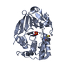
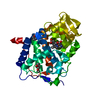
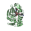
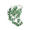
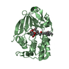
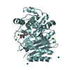
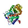
| Title | Crystal structure of a Protein Phosphatase-1: Calcineurin Hybrid Bound to Okadaic Acid | ||||||
 Components Components | Serine/threonine protein phosphatase PP1-gamma catalytic subunit Serine/threonine-specific protein kinase Serine/threonine-specific protein kinase | ||||||
 Keywords Keywords | HYDROLASE | ||||||
| Function / homology |  Function and homology information Function and homology informationPTW/PP1 phosphatase complex / regulation of nucleocytoplasmic transport / protein phosphatase 1 binding / lamin binding / SHOC2 M1731 mutant abolishes MRAS complex function / Gain-of-function MRAS complexes activate RAF signaling / microtubule organizing center / myosin phosphatase activity / protein serine/threonine phosphatase activity / glycogen metabolic process ...PTW/PP1 phosphatase complex / regulation of nucleocytoplasmic transport / protein phosphatase 1 binding / lamin binding / SHOC2 M1731 mutant abolishes MRAS complex function / Gain-of-function MRAS complexes activate RAF signaling / microtubule organizing center / myosin phosphatase activity / protein serine/threonine phosphatase activity / glycogen metabolic process / protein-serine/threonine phosphatase / entrainment of circadian clock by photoperiod / Triglyceride catabolism / phosphatase activity / phosphoprotein phosphatase activity / cleavage furrow / blastocyst development / Amplification of signal from unattached kinetochores via a MAD2 inhibitory signal / Mitotic Prometaphase / EML4 and NUDC in mitotic spindle formation / positive regulation of glial cell proliferation / Resolution of Sister Chromatid Cohesion / protein dephosphorylation / Downregulation of TGF-beta receptor signaling / RHO GTPases Activate Formins / RAF activation / circadian regulation of gene expression / regulation of circadian rhythm / neuron differentiation / kinetochore / Separation of Sister Chromatids / MAPK cascade / Circadian Clock / presynapse / midbody / spermatogenesis / mitochondrial outer membrane / dendritic spine / nuclear speck / cell cycle / cell division / protein domain specific binding / focal adhesion / glutamatergic synapse / protein-containing complex binding / nucleolus / protein kinase binding / protein-containing complex / mitochondrion / RNA binding / metal ion binding / nucleus / cytosol / cytoplasmSimilarity search - Function | ||||||
| Biological species |  Homo sapiens (human) Homo sapiens (human) | ||||||
| Method | X-RAY DIFFRACTION / MOLECULAR REPLACEMENT / Resolution: 2 Å | ||||||
 Authors Authors | Maynes, J.T. / Perreault, K.R. / Cherney, M.M. / Luu, H.A. / James, M.N.G. / Holmes, C.F.B. | ||||||
 Citation Citation | Journal: J.Biol.Chem. / Year: 2004 Title: Crystal Structure and Mutagenesis of a Protein Phosphatase-1:Calcineurin Hybrid Elucidate the Role of the {beta}12-{beta}13 Loop in Inhibitor Binding Authors: Maynes, J.T. / Perreault, K.R. / Cherney, M.M. / Luu, H.A. / James, M.N.G. / Holmes, C.F.B. | ||||||
| History |
|
- Structure visualization
| Structure viewer | Molecule: MolmilJmol/JSmol |
|---|
- Downloads & links
-Download
| PDBx/mmCIF format | 1u32.cif.gz | 78.8 KB | Display | PDBx/mmCIF format |
|---|---|---|---|---|
| PDB format | pdb1u32.ent.gz | 57.3 KB | Display | PDB format |
| PDBx/mmJSON format | 1u32.json.gz | Tree view | PDBx/mmJSON format | |
| Others |  Other downloads Other downloads |
-Validation report
| Arichive directory | https://data.pdbj.org/pub/pdb/validation_reports/u3/1u32ftp://data.pdbj.org/pub/pdb/validation_reports/u3/1u32 | HTTPS FTP |
|---|
-Related structure data
| Related structure data | 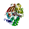 1jk7S S: Starting model for refinement |
|---|---|
| Similar structure data |


-Links
 PDBj
PDBj


- Assembly
| Deposited unit | 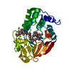
| ||||||||
|---|---|---|---|---|---|---|---|---|---|
| 1 |
| ||||||||
| Unit cell |
|
-Components
| #1: Protein | Serine/threonine-specific protein kinase / Protein Phosphatase-1: Calcineurin hybrid / PP-1G / Protein phosphatase 1C catalytic subunit Mass: 33771.910 Da / Num. of mol.: 1 / Fragment: residues 6-298 Source method: isolated from a genetically manipulated source Source: (gene. exp.) Homo sapiens (human) / Gene: PPP1CC / Plasmid: pet / Production host:  Escherichia coli (E. coli) Escherichia coli (E. coli)References: UniProt: P36873, protein-serine/threonine phosphatase | ||||||
|---|---|---|---|---|---|---|---|
| #2: Chemical |   Mass: 54.938 Da / Num. of mol.: 2 / Source method: obtained synthetically / Formula: Mn Mass: 54.938 Da / Num. of mol.: 2 / Source method: obtained synthetically / Formula: Mn#3: Chemical | ChemComp-OKA / | Okadaic acid  Mass: 805.003 Da / Num. of mol.: 1 / Source method: obtained synthetically / Formula: C44H68O13 / Comment: inhibitor, toxin*YM Mass: 805.003 Da / Num. of mol.: 1 / Source method: obtained synthetically / Formula: C44H68O13 / Comment: inhibitor, toxin*YM#4: Chemical | 2-Mercaptoethanol  Mass: 78.133 Da / Num. of mol.: 3 / Source method: obtained synthetically / Formula: C2H6OS Mass: 78.133 Da / Num. of mol.: 3 / Source method: obtained synthetically / Formula: C2H6OS#5: Water | ChemComp-HOH / | Water Mass: 18.015 Da / Num. of mol.: 89 / Source method: isolated from a natural source / Formula: H2O Mass: 18.015 Da / Num. of mol.: 89 / Source method: isolated from a natural source / Formula: H2O |
-Experimental details
-Experiment
| Experiment | Method: X-RAY DIFFRACTION / Number of used crystals: 1 |
|---|
- Sample preparation
| Crystal | Density Matthews: 2.4 Å3/Da / Density % sol: 47 % |
|---|---|
| Crystal grow | Temperature: 298 K / Method: vapor diffusion, hanging drop / pH: 8 Details: lithium sulfate, tris, PEG 400, mercaptoethanol, pH 8.0, VAPOR DIFFUSION, HANGING DROP, temperature 298K |
-Data collection
| Diffraction | Mean temperature: 100 K |
|---|---|
| Diffraction source | Source: ROTATING ANODE / Type: RIGAKU RUH3R / Wavelength: 1.5418 |
| Detector | Type: RIGAKU RAXIS IV / Detector: IMAGE PLATE / Date: Jul 22, 2002 / Details: osmic mirrors |
| Radiation | Monochromator: mirrors / Protocol: SINGLE WAVELENGTH / Monochromatic (M) / Laue (L): M / Scattering type: x-ray |
| Radiation wavelength | Wavelength: 1.5418 Å / Relative weight: 1 |
| Reflection | Resolution: 2→40 Å / Num. all: 21891 / Num. obs: 21304 / % possible obs: 97.3 % / Observed criterion σ(F): 1.9 / Observed criterion σ(I): 2.5 / Redundancy: 5.8 % / Biso Wilson estimate: 15 Å2 / Rmerge(I) obs: 0.076 / Rsym value: 0.057 / Net I/σ(I): 21.1 |
| Reflection shell | Resolution: 2→2.07 Å / Redundancy: 5.7 % / Rmerge(I) obs: 0.369 / Mean I/σ(I) obs: 5.02 / Num. unique all: 2063 / Rsym value: 0.36 / % possible all: 99 |
- Processing
| Software |
| ||||||||||||||||||||||||||||||||||||
|---|---|---|---|---|---|---|---|---|---|---|---|---|---|---|---|---|---|---|---|---|---|---|---|---|---|---|---|---|---|---|---|---|---|---|---|---|---|
| Refinement | Method to determine structure: MOLECULAR REPLACEMENT Starting model: PDB ID 1JK7 Resolution: 2→36.01 Å / Rfactor Rfree error: 0.007 / Data cutoff high absF: 1946384.85 / Data cutoff low absF: 0 / Isotropic thermal model: RESTRAINED / Cross valid method: THROUGHOUT / σ(F): 0 / Stereochemistry target values: Engh & Huber
| ||||||||||||||||||||||||||||||||||||
| Solvent computation | Solvent model: FLAT MODEL / Bsol: 37.2487 Å2 / ksol: 0.359391 e/Å3 | ||||||||||||||||||||||||||||||||||||
| Displacement parameters | Biso mean: 27.3 Å2
| ||||||||||||||||||||||||||||||||||||
| Refine analyze |
| ||||||||||||||||||||||||||||||||||||
| Refinement step | Cycle: LAST / Resolution: 2→36.01 Å
| ||||||||||||||||||||||||||||||||||||
| Refine LS restraints |
| ||||||||||||||||||||||||||||||||||||
| LS refinement shell | Resolution: 2→2.13 Å / Rfactor Rfree error: 0.019 / Total num. of bins used: 6
| ||||||||||||||||||||||||||||||||||||
| Xplor file |
|
