 Movie
Movie Controller

[English] 日本語
 Yorodumi
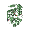
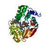
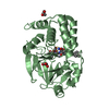
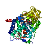
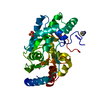
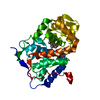
- PDB-4ut3: X-ray structure of the human PP1 gamma catalytic subunit treated ... -
+ Open data

- Basic information
| Entry | Database: PDB / ID: 4ut3 | ||||||
|---|---|---|---|---|---|---|---|
| Title | X-ray structure of the human PP1 gamma catalytic subunit treated with hydrogen peroxide | ||||||
 Components Components | (SERINE/THREONINE-PROTEIN PHOSPHATASE PP1-GAMMA CATALYTIC SUBUNIT) x 2 | ||||||
 Keywords Keywords |  HYDROLASE / METAL CENTER / METALLOPROTEIN / ENZYME ACTIVATION / PHOSPHOPROTEIN PHOSPHATASES / PROTEIN PHOSPHATASE 1 HYDROLASE / METAL CENTER / METALLOPROTEIN / ENZYME ACTIVATION / PHOSPHOPROTEIN PHOSPHATASES / PROTEIN PHOSPHATASE 1 | ||||||
| Function / homology |  Function and homology information Function and homology informationPTW/PP1 phosphatase complex / regulation of nucleocytoplasmic transport / protein phosphatase 1 binding / lamin binding / SHOC2 M1731 mutant abolishes MRAS complex function / Gain-of-function MRAS complexes activate RAF signaling / microtubule organizing center / myosin phosphatase activity / protein serine/threonine phosphatase activity / glycogen metabolic process ...PTW/PP1 phosphatase complex / regulation of nucleocytoplasmic transport / protein phosphatase 1 binding / lamin binding / SHOC2 M1731 mutant abolishes MRAS complex function / Gain-of-function MRAS complexes activate RAF signaling / microtubule organizing center / myosin phosphatase activity / protein serine/threonine phosphatase activity / glycogen metabolic process / protein-serine/threonine phosphatase / entrainment of circadian clock by photoperiod / Triglyceride catabolism / phosphatase activity / phosphoprotein phosphatase activity / cleavage furrow / blastocyst development / Amplification of signal from unattached kinetochores via a MAD2 inhibitory signal / Mitotic Prometaphase / EML4 and NUDC in mitotic spindle formation / positive regulation of glial cell proliferation / Resolution of Sister Chromatid Cohesion / protein dephosphorylation / Downregulation of TGF-beta receptor signaling / RHO GTPases Activate Formins / RAF activation / circadian regulation of gene expression / regulation of circadian rhythm / neuron differentiation / kinetochore / Separation of Sister Chromatids / MAPK cascade / Circadian Clock / presynapse / midbody / spermatogenesis / mitochondrial outer membrane / dendritic spine / nuclear speck / cell cycle / cell division / protein domain specific binding / focal adhesion / glutamatergic synapse / protein-containing complex binding / nucleolus / protein kinase binding / protein-containing complex / mitochondrion / RNA binding / metal ion binding / nucleus / cytosol / cytoplasmSimilarity search - Function | ||||||
| Biological species |  HOMO SAPIENS (human) HOMO SAPIENS (human) | ||||||
| Method | X-RAY DIFFRACTION / SYNCHROTRON / MOLECULAR REPLACEMENT / Resolution: 2.19 Å | ||||||
 Authors Authors | Zeh Silva, M. / Kopec, J. / Fotinou, D. / Steiner, R.A. | ||||||
 Citation Citation | Journal: Embo J. / Year: 2016 Title: Targeted Redox Inhibition of Protein Phosphatase 1 by Nox4 Regulates Eif2Alpha-Mediated Stress Signaling. Authors: Santos, C.X. / Hafstad, A.D. / Beretta, M. / Zhang, M. / Molenaar, C. / Kopec, J. / Fotinou, D. / Murray, T.V. / Cobb, A.M. / Martin, D. / Zeh Silva, M. / Anilkumar, N. / Schroder, K. / ...Authors: Santos, C.X. / Hafstad, A.D. / Beretta, M. / Zhang, M. / Molenaar, C. / Kopec, J. / Fotinou, D. / Murray, T.V. / Cobb, A.M. / Martin, D. / Zeh Silva, M. / Anilkumar, N. / Schroder, K. / Shanahan, C.M. / Brewer, A.C. / Brandes, R.P. / Blanc, E. / Parsons, M. / Belousov, V. / Cammack, R. / Hider, R.C. / Steiner, R.A. / Shah, A.M. | ||||||
| History |
|
- Structure visualization
| Structure viewer | Molecule: MolmilJmol/JSmol |
|---|
- Downloads & links
-Download
| PDBx/mmCIF format | 4ut3.cif.gz | 136 KB | Display | PDBx/mmCIF format |
|---|---|---|---|---|
| PDB format | pdb4ut3.ent.gz | 106.1 KB | Display | PDB format |
| PDBx/mmJSON format | 4ut3.json.gz | Tree view | PDBx/mmJSON format | |
| Others |  Other downloads Other downloads |
-Validation report
| Arichive directory | https://data.pdbj.org/pub/pdb/validation_reports/ut/4ut3ftp://data.pdbj.org/pub/pdb/validation_reports/ut/4ut3 | HTTPS FTP |
|---|
-Related structure data
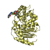
| Related structure data |  4ut2C 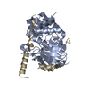 2o8aS C: citing same article ( S: Starting model for refinement |
|---|---|
| Similar structure data |


-Links
 PDBj
PDBj


- Assembly
| Deposited unit | 
| ||||||||
|---|---|---|---|---|---|---|---|---|---|
| 1 | 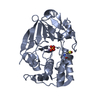
| ||||||||
| 2 | 
| ||||||||
| Unit cell |
|
-Components
| #1: Protein | Mass: 37046.777 Da / Num. of mol.: 1 Source method: isolated from a genetically manipulated source Source: (gene. exp.) HOMO SAPIENS (human) / Production host:  ESCHERICHIA COLI DH5[ALPHA] (bacteria) ESCHERICHIA COLI DH5[ALPHA] (bacteria)References: UniProt: P36873, protein-serine/threonine phosphatase | ||||
|---|---|---|---|---|---|
| #2: Protein | Mass: 37062.777 Da / Num. of mol.: 1 Source method: isolated from a genetically manipulated source Source: (gene. exp.) HOMO SAPIENS (human) / Production host: ESCHERICHIA COLI DH5[ALPHA] (bacteria)References: UniProt: P36873, protein-serine/threonine phosphatase | ||||
| #3: Chemical | ChemComp-MN /   Mass: 54.938 Da / Num. of mol.: 4 / Source method: obtained synthetically / Formula: Mn Mass: 54.938 Da / Num. of mol.: 4 / Source method: obtained synthetically / Formula: Mn#4: Chemical | Phosphate  Mass: 94.971 Da / Num. of mol.: 2 / Source method: obtained synthetically / Formula: PO4 Mass: 94.971 Da / Num. of mol.: 2 / Source method: obtained synthetically / Formula: PO4#5: Water | ChemComp-HOH / | Water Mass: 18.015 Da / Num. of mol.: 161 / Source method: isolated from a natural source / Formula: H2O Mass: 18.015 Da / Num. of mol.: 161 / Source method: isolated from a natural source / Formula: H2O |
-Experimental details
-Experiment
| Experiment | Method: X-RAY DIFFRACTION / Number of used crystals: 1 |
|---|
- Sample preparation
| Crystal | Density Matthews: 2.46 Å3/Da / Density % sol: 49.96 % / Description: NONE |
|---|---|
| Crystal grow | pH: 9 Details: 7-12% PEG3350, 0.1 M BICINE, PH 9.0 SOAKING IN RESERVOIR ENRICHED WITH 50MM H2O2 FOR 10 MINS CRYOPROTECTION IN RESEVOIR ENRICHED WITH 25% MPD |
-Data collection
| Diffraction | Mean temperature: 100 K |
|---|---|
| Diffraction source | Source: SYNCHROTRON / Site: Diamond  / Beamline: I03 / Wavelength: 0.9762 / Beamline: I03 / Wavelength: 0.9762 |
| Detector | Type: DECTRIS PILATUS 6M / Detector: PIXEL / Date: Dec 19, 2012 |
| Radiation | Protocol: SINGLE WAVELENGTH / Monochromatic (M) / Laue (L): M / Scattering type: x-ray |
| Radiation wavelength | Wavelength: 0.9762 Å / Relative weight: 1 |
| Reflection | Resolution: 2.2→45.4 Å / Num. obs: 35328 / % possible obs: 97.5 % / Observed criterion σ(I): -1 / Redundancy: 3.3 % / Rmerge(I) obs: 0.08 / Net I/σ(I): 7.9 |
| Reflection shell | Resolution: 2.2→2.26 Å / Redundancy: 3.3 % / Rmerge(I) obs: 0.62 / Mean I/σ(I) obs: 2.1 / % possible all: 97.3 |
- Processing
| Software |
| ||||||||||||||||||||||||||||||||||||||||||||||||||||||||||||||||||||||||||||||||||||||||||||||||||||||||||||||||||||||||||||||||||||||||||||||||||||||||||||||||||||||||||||||||||||||
|---|---|---|---|---|---|---|---|---|---|---|---|---|---|---|---|---|---|---|---|---|---|---|---|---|---|---|---|---|---|---|---|---|---|---|---|---|---|---|---|---|---|---|---|---|---|---|---|---|---|---|---|---|---|---|---|---|---|---|---|---|---|---|---|---|---|---|---|---|---|---|---|---|---|---|---|---|---|---|---|---|---|---|---|---|---|---|---|---|---|---|---|---|---|---|---|---|---|---|---|---|---|---|---|---|---|---|---|---|---|---|---|---|---|---|---|---|---|---|---|---|---|---|---|---|---|---|---|---|---|---|---|---|---|---|---|---|---|---|---|---|---|---|---|---|---|---|---|---|---|---|---|---|---|---|---|---|---|---|---|---|---|---|---|---|---|---|---|---|---|---|---|---|---|---|---|---|---|---|---|---|---|---|---|
| Refinement | Method to determine structure: MOLECULAR REPLACEMENT Starting model: PDB ENTRY 2O8A Resolution: 2.19→45.4 Å / Cor.coef. Fo:Fc: 0.966 / Cor.coef. Fo:Fc free: 0.947 / SU B: 4.46 / SU ML: 0.113 / Cross valid method: THROUGHOUT / ESU R: 0.05 / ESU R Free: 0.039 / Stereochemistry target values: MAXIMUM LIKELIHOOD Details: HYDROGENS HAVE BEEN ADDED IN THE RIDING POSITIONS. U VALUES REFINED INDIVIDUALLY
| ||||||||||||||||||||||||||||||||||||||||||||||||||||||||||||||||||||||||||||||||||||||||||||||||||||||||||||||||||||||||||||||||||||||||||||||||||||||||||||||||||||||||||||||||||||||
| Solvent computation | Ion probe radii: 0.8 Å / Shrinkage radii: 0.8 Å / VDW probe radii: 1.2 Å / Solvent model: MASK | ||||||||||||||||||||||||||||||||||||||||||||||||||||||||||||||||||||||||||||||||||||||||||||||||||||||||||||||||||||||||||||||||||||||||||||||||||||||||||||||||||||||||||||||||||||||
| Displacement parameters | Biso mean: 47.211 Å2
| ||||||||||||||||||||||||||||||||||||||||||||||||||||||||||||||||||||||||||||||||||||||||||||||||||||||||||||||||||||||||||||||||||||||||||||||||||||||||||||||||||||||||||||||||||||||
| Refinement step | Cycle: LAST / Resolution: 2.19→45.4 Å
| ||||||||||||||||||||||||||||||||||||||||||||||||||||||||||||||||||||||||||||||||||||||||||||||||||||||||||||||||||||||||||||||||||||||||||||||||||||||||||||||||||||||||||||||||||||||
| Refine LS restraints |
|