 Movie
Movie Controller

[English] 日本語
 Yorodumi
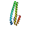
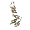
- PDB-1s94: Crystal structure of the Habc domain of neuronal syntaxin from th... -
+ Open data

- Basic information
| Entry | Database: PDB / ID: 1s94 | ||||||
|---|---|---|---|---|---|---|---|
| Title | Crystal structure of the Habc domain of neuronal syntaxin from the squid Loligo pealei | ||||||
 Components Components | s-syntaxin | ||||||
 Keywords Keywords | ENDOCYTOSIS/EXOCYTOSIS /  three helix bundle / structural plasticity / ENDOCYTOSIS-EXOCYTOSIS COMPLEX three helix bundle / structural plasticity / ENDOCYTOSIS-EXOCYTOSIS COMPLEX | ||||||
| Function / homology |  Function and homology informationSNAP receptor activity / vesicle-mediated transport / intracellular protein transport / membrane Function and homology informationSNAP receptor activity / vesicle-mediated transport / intracellular protein transport / membraneSimilarity search - Function | ||||||
| Biological species |  Loligo pealei (longfin inshore squid) Loligo pealei (longfin inshore squid) | ||||||
| Method | X-RAY DIFFRACTION / SYNCHROTRON / MOLECULAR REPLACEMENT / Resolution: 3.34 Å | ||||||
 Authors Authors | Bracher, A. / Weissenhorn, W. | ||||||
 Citation Citation | Journal: BMC STRUCT.BIOL. / Year: 2004 Title: Crystal structure of the Habc domain of neuronal syntaxin from the squid Loligo pealei reveals conformational plasticity at its C-terminus Authors: Bracher, A. / Weissenhorn, W. | ||||||
| History |
|
- Structure visualization
| Structure viewer | Molecule: MolmilJmol/JSmol |
|---|
- Downloads & links
-Download
| PDBx/mmCIF format | 1s94.cif.gz | 56.3 KB | Display | PDBx/mmCIF format |
|---|---|---|---|---|
| PDB format | pdb1s94.ent.gz | 39.8 KB | Display | PDB format |
| PDBx/mmJSON format | 1s94.json.gz | Tree view | PDBx/mmJSON format | |
| Others |  Other downloads Other downloads |
-Validation report
| Arichive directory | https://data.pdbj.org/pub/pdb/validation_reports/s9/1s94ftp://data.pdbj.org/pub/pdb/validation_reports/s9/1s94 | HTTPS FTP |
|---|
-Related structure data
| Related structure data | 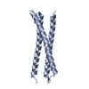 1ez3S S: Starting model for refinement |
|---|---|
| Similar structure data |


-Links
 PDBj
PDBj
- Assembly
| Deposited unit | 
| ||||||||
|---|---|---|---|---|---|---|---|---|---|
| 1 | 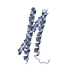
| ||||||||
| 2 | 
| ||||||||
| Unit cell |
|
-Components
| #1: Protein | Mass: 20715.221 Da / Num. of mol.: 2 / Fragment: Habc domain / Mutation: M1R Source method: isolated from a genetically manipulated source Source: (gene. exp.) Loligo pealei (longfin inshore squid) / Plasmid: pQE30 / Production host:  Escherichia coli (E. coli) / Strain (production host): Bl21 pUBS / References: UniProt: O46345 Escherichia coli (E. coli) / Strain (production host): Bl21 pUBS / References: UniProt: O46345 |
|---|
-Experimental details
-Experiment
| Experiment | Method: X-RAY DIFFRACTION / Number of used crystals: 1 |
|---|
- Sample preparation
| Crystal | Density Matthews: 3.74 Å3/Da / Density % sol: 66.9 % |
|---|---|
| Crystal grow | Temperature: 293 K / Method: vapor diffusion, hanging drop / pH: 8.5 Details: 30% PEG 400, 0.2M Na-citrate, 0.1M Tris HCl, 20mM HEPES-KOH, 100mM KCl, pH 8.5, VAPOR DIFFUSION, HANGING DROP, temperature 293K |
-Data collection
| Diffraction | Mean temperature: 100 K |
|---|---|
| Diffraction source | Source: SYNCHROTRON / Site: ESRF  / Beamline: ID29 / Wavelength: 1.0088 Å / Beamline: ID29 / Wavelength: 1.0088 Å |
| Detector | Type: ADSC QUANTUM 4 / Detector: CCD / Date: Jun 25, 2001 |
| Radiation | Protocol: SINGLE WAVELENGTH / Monochromatic (M) / Laue (L): M / Scattering type: x-ray |
| Radiation wavelength | Wavelength: 1.0088 Å / Relative weight: 1 |
| Reflection | Resolution: 3.3→15 Å / Num. all: 9361 / Num. obs: 9304 / % possible obs: 99.4 % / Observed criterion σ(F): 0 / Observed criterion σ(I): 0 / Redundancy: 6.4 % / Rmerge(I) obs: 0.074 / Net I/σ(I): 14.8 |
| Reflection shell | Resolution: 3.3→3.42 Å / Rmerge(I) obs: 0.375 / Mean I/σ(I) obs: 4.6 / Num. unique all: 906 / % possible all: 100 |
- Processing
| Software |
| ||||||||||||||||||||||||||||||||||||||||||||||||||||||||||||||||||||||||||||||||||||||||||||||||||||||||||||||||||||||||||||||||||||||||||||||||||||||
|---|---|---|---|---|---|---|---|---|---|---|---|---|---|---|---|---|---|---|---|---|---|---|---|---|---|---|---|---|---|---|---|---|---|---|---|---|---|---|---|---|---|---|---|---|---|---|---|---|---|---|---|---|---|---|---|---|---|---|---|---|---|---|---|---|---|---|---|---|---|---|---|---|---|---|---|---|---|---|---|---|---|---|---|---|---|---|---|---|---|---|---|---|---|---|---|---|---|---|---|---|---|---|---|---|---|---|---|---|---|---|---|---|---|---|---|---|---|---|---|---|---|---|---|---|---|---|---|---|---|---|---|---|---|---|---|---|---|---|---|---|---|---|---|---|---|---|---|---|---|---|---|
| Refinement | Method to determine structure: MOLECULAR REPLACEMENT Starting model: pdb entry 1EZ3, chain A Resolution: 3.34→14.97 Å / Cor.coef. Fo:Fc: 0.847 / Cor.coef. Fo:Fc free: 0.818 / SU B: 25.633 / SU ML: 0.43 / TLS residual ADP flag: LIKELY RESIDUAL / Cross valid method: THROUGHOUT / σ(F): 0 / ESU R: 0.889 / ESU R Free: 0.55 / Stereochemistry target values: MAXIMUM LIKELIHOOD / Details: HYDROGENS HAVE BEEN ADDED IN THE RIDING POSITIONS
| ||||||||||||||||||||||||||||||||||||||||||||||||||||||||||||||||||||||||||||||||||||||||||||||||||||||||||||||||||||||||||||||||||||||||||||||||||||||
| Solvent computation | Ion probe radii: 0.8 Å / Shrinkage radii: 0.8 Å / VDW probe radii: 1.4 Å / Solvent model: BABINET MODEL WITH MASK | ||||||||||||||||||||||||||||||||||||||||||||||||||||||||||||||||||||||||||||||||||||||||||||||||||||||||||||||||||||||||||||||||||||||||||||||||||||||
| Displacement parameters | Biso mean: 42.344 Å2
| ||||||||||||||||||||||||||||||||||||||||||||||||||||||||||||||||||||||||||||||||||||||||||||||||||||||||||||||||||||||||||||||||||||||||||||||||||||||
| Refinement step | Cycle: LAST / Resolution: 3.34→14.97 Å
| ||||||||||||||||||||||||||||||||||||||||||||||||||||||||||||||||||||||||||||||||||||||||||||||||||||||||||||||||||||||||||||||||||||||||||||||||||||||
| Refine LS restraints |
| ||||||||||||||||||||||||||||||||||||||||||||||||||||||||||||||||||||||||||||||||||||||||||||||||||||||||||||||||||||||||||||||||||||||||||||||||||||||
| LS refinement shell | Resolution: 3.34→3.421 Å / Total num. of bins used: 20 /
| ||||||||||||||||||||||||||||||||||||||||||||||||||||||||||||||||||||||||||||||||||||||||||||||||||||||||||||||||||||||||||||||||||||||||||||||||||||||
| Refinement TLS params. | Method: refined / Refine-ID: X-RAY DIFFRACTION
| ||||||||||||||||||||||||||||||||||||||||||||||||||||||||||||||||||||||||||||||||||||||||||||||||||||||||||||||||||||||||||||||||||||||||||||||||||||||
| Refinement TLS group |
|
