 Movie
Movie Controller


+ Open data

- Basic information
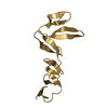
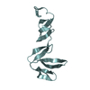
| Entry | Database: PDB / ID: 4c8w | ||||||
|---|---|---|---|---|---|---|---|
| Title | Xenopus RSPO2 Fu1-Fu2 crystal form II | ||||||
 Components Components | R-SPONDIN-2 | ||||||
 Keywords Keywords |  SIGNALING PROTEIN / WNT / ZNRF3 / RNF43 / LGR4 / LGR5 / LGR6 / RSPO / R-SPO / RSPO1 / RSPO3 / RSPO4 / RECEPTOR / MEMBRANE / SIGNALLING SIGNALING PROTEIN / WNT / ZNRF3 / RNF43 / LGR4 / LGR5 / LGR6 / RSPO / R-SPO / RSPO1 / RSPO3 / RSPO4 / RECEPTOR / MEMBRANE / SIGNALLING | ||||||
| Function / homology |  Function and homology information Function and homology informationanal fin development / caudal fin development / dorsal fin development / pectoral fin development / pelvic fin development / Regulation of FZD by ubiquitination / skeletal system development / Wnt signaling pathway / heparin binding / extracellular regionSimilarity search - Function | ||||||
| Biological species | XENOPUS TROPICALIS | ||||||
| Method | X-RAY DIFFRACTION / SYNCHROTRON / OTHER / Resolution: 3.1 Å | ||||||
 Authors Authors | Zebisch, M. / Jones, E.Y. | ||||||
 Citation Citation | Journal: Nat.Commun. / Year: 2013 Title: Structural and Molecular Basis of Znrf3/Rnf43 Transmembrane Ubiquitin Ligase Inhibition by the Wnt Agonist R-Spondin. Authors: Zebisch, M. / Xu, Y. / Krastev, C. / Macdonald, B.T. / Chen, M. / Gilbert, R.J.C. / He, X. / Jones, E.Y. | ||||||
| History |
|
- Structure visualization
| Structure viewer | Molecule: MolmilJmol/JSmol |
|---|
- Downloads & links
-Download
| PDBx/mmCIF format | 4c8w.cif.gz | 47.3 KB | Display | PDBx/mmCIF format |
|---|---|---|---|---|
| PDB format | pdb4c8w.ent.gz | 37.6 KB | Display | PDB format |
| PDBx/mmJSON format | 4c8w.json.gz | Tree view | PDBx/mmJSON format | |
| Others |  Other downloads Other downloads |
-Validation report
| Arichive directory | https://data.pdbj.org/pub/pdb/validation_reports/c8/4c8wftp://data.pdbj.org/pub/pdb/validation_reports/c8/4c8w | HTTPS FTP |
|---|
-Related structure data
| Related structure data | 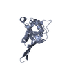 4c84C 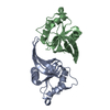 4c85C 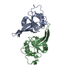 4c86C  4c8aC 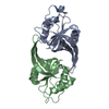 4c8cC 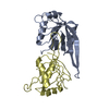 4c8fC 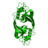 4c8pC 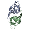 4c8tC 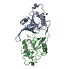 4c8uC 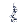 4c8vC 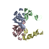 4c99C 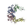 4c9aC  4c9eC 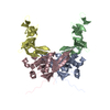 4c9rC 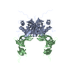 4c9uC 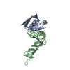 4c9vC C: citing same article ( |
|---|---|
| Similar structure data |


-Links
 PDBj
PDBj


- Assembly
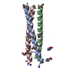
| Deposited unit | 
| ||||||||
|---|---|---|---|---|---|---|---|---|---|
| 1 | 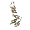
| ||||||||
| 2 | 
| ||||||||
| Unit cell |
|
-Components
| #1: Protein | Mass: 13781.749 Da / Num. of mol.: 2 / Fragment: FU1-FU2, RESIDUES 35-144 Source method: isolated from a genetically manipulated source Source: (gene. exp.) XENOPUS (SILURANA) TROPICALIS (tropical clawed frog)Plasmid: PHLSEC / Cell line (production host): HEK293T / Production host:  HOMO SAPIENS (human) / References: UniProt: Q5M7L6 HOMO SAPIENS (human) / References: UniProt: Q5M7L6 |
|---|
-Experimental details
-Experiment
| Experiment | Method: X-RAY DIFFRACTION |
|---|
- Sample preparation
| Crystal | Density Matthews: 2.65 Å3/Da / Density % sol: 53.56 % / Description: NONE |
|---|
-Data collection
| Diffraction | Mean temperature: 100 K |
|---|---|
| Diffraction source | Source: SYNCHROTRON / Site: Diamond  / Beamline: I02 / Wavelength: 0.9795 / Beamline: I02 / Wavelength: 0.9795 |
| Detector | Type: DECTRIS PIXEL / Detector: PIXEL / Date: May 31, 2012 |
| Radiation | Protocol: SINGLE WAVELENGTH / Monochromatic (M) / Laue (L): M / Scattering type: x-ray |
| Radiation wavelength | Wavelength: 0.9795 Å / Relative weight: 1 |
| Reflection | Resolution: 3.1→87 Å / Num. obs: 5686 / % possible obs: 99.1 % / Observed criterion σ(I): -3 / Redundancy: 4.8 % / Rmerge(I) obs: 0.19 / Net I/σ(I): 6.8 |
- Processing
| Software | Name: REFMAC / Version: 5.7.0029 / Classification: refinement | ||||||||||||||||||||||||||||||||||||||||||||||||||||||||||||||||||||||||||||||||||||||||||||||||||||||||||||||||||||||||||||||||||||||||||||||||||||||||||||||||||||||||||||||||||||||
|---|---|---|---|---|---|---|---|---|---|---|---|---|---|---|---|---|---|---|---|---|---|---|---|---|---|---|---|---|---|---|---|---|---|---|---|---|---|---|---|---|---|---|---|---|---|---|---|---|---|---|---|---|---|---|---|---|---|---|---|---|---|---|---|---|---|---|---|---|---|---|---|---|---|---|---|---|---|---|---|---|---|---|---|---|---|---|---|---|---|---|---|---|---|---|---|---|---|---|---|---|---|---|---|---|---|---|---|---|---|---|---|---|---|---|---|---|---|---|---|---|---|---|---|---|---|---|---|---|---|---|---|---|---|---|---|---|---|---|---|---|---|---|---|---|---|---|---|---|---|---|---|---|---|---|---|---|---|---|---|---|---|---|---|---|---|---|---|---|---|---|---|---|---|---|---|---|---|---|---|---|---|---|---|
| Refinement | Method to determine structure: OTHER Starting model: NONE Resolution: 3.1→87.12 Å / Cor.coef. Fo:Fc: 0.885 / Cor.coef. Fo:Fc free: 0.837 / SU B: 23.074 / SU ML: 0.403 / Cross valid method: THROUGHOUT / ESU R Free: 0.529 / Stereochemistry target values: MAXIMUM LIKELIHOOD Details: HYDROGENS HAVE BEEN ADDED IN THE RIDING POSITIONS. U VALUES REFINED INDIVIDUALLY.
| ||||||||||||||||||||||||||||||||||||||||||||||||||||||||||||||||||||||||||||||||||||||||||||||||||||||||||||||||||||||||||||||||||||||||||||||||||||||||||||||||||||||||||||||||||||||
| Solvent computation | Ion probe radii: 0.8 Å / Shrinkage radii: 0.8 Å / VDW probe radii: 1.2 Å / Solvent model: MASK | ||||||||||||||||||||||||||||||||||||||||||||||||||||||||||||||||||||||||||||||||||||||||||||||||||||||||||||||||||||||||||||||||||||||||||||||||||||||||||||||||||||||||||||||||||||||
| Displacement parameters | Biso mean: 57.287 Å2
| ||||||||||||||||||||||||||||||||||||||||||||||||||||||||||||||||||||||||||||||||||||||||||||||||||||||||||||||||||||||||||||||||||||||||||||||||||||||||||||||||||||||||||||||||||||||
| Refinement step | Cycle: LAST / Resolution: 3.1→87.12 Å
| ||||||||||||||||||||||||||||||||||||||||||||||||||||||||||||||||||||||||||||||||||||||||||||||||||||||||||||||||||||||||||||||||||||||||||||||||||||||||||||||||||||||||||||||||||||||
| Refine LS restraints |
|