 Movie
Movie Controller

[English] 日本語
 Yorodumi
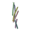
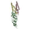
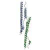
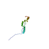
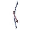
- PDB-1l4a: X-RAY STRUCTURE OF THE NEURONAL COMPLEXIN/SNARE COMPLEX FROM THE ... -
+ Open data

- Basic information
| Entry | Database: PDB / ID: 1l4a | ||||||
|---|---|---|---|---|---|---|---|
| Title | X-RAY STRUCTURE OF THE NEURONAL COMPLEXIN/SNARE COMPLEX FROM THE SQUID LOLIGO PEALEI | ||||||
 Components Components |
| ||||||
 Keywords Keywords | ENDOCYTOSIS/EXOCYTOSIS /  SNARE / SNARE complex / membrane fusion / neurotransmission / ENDOCYTOSIS-EXOCYTOSIS COMPLEX SNARE / SNARE complex / membrane fusion / neurotransmission / ENDOCYTOSIS-EXOCYTOSIS COMPLEX | ||||||
| Function / homology |  Function and homology informationSNAP receptor activity / neurotransmitter transport / syntaxin binding / exocytosis / vesicle-mediated transport / intracellular protein transport / synaptic vesicle membrane / membrane => GO:0016020 / neuron projection / synapse ...SNAP receptor activity / neurotransmitter transport / syntaxin binding / exocytosis / vesicle-mediated transport / intracellular protein transport / synaptic vesicle membrane / membrane => GO:0016020 / neuron projection / synapse / membrane / cytosol Function and homology informationSNAP receptor activity / neurotransmitter transport / syntaxin binding / exocytosis / vesicle-mediated transport / intracellular protein transport / synaptic vesicle membrane / membrane => GO:0016020 / neuron projection / synapse ...SNAP receptor activity / neurotransmitter transport / syntaxin binding / exocytosis / vesicle-mediated transport / intracellular protein transport / synaptic vesicle membrane / membrane => GO:0016020 / neuron projection / synapse / membrane / cytosolSimilarity search - Function | ||||||
| Biological species |  Loligo pealei (longfin inshore squid) Loligo pealei (longfin inshore squid) | ||||||
| Method | X-RAY DIFFRACTION / SYNCHROTRON / MOLECULAR REPLACEMENT / Resolution: 2.95 Å | ||||||
 Authors Authors | Bracher, A. / Kadlec, J. / Betz, H. / Weissenhorn, W. | ||||||
 Citation Citation | Journal: J.Biol.Chem. / Year: 2002 Title: X-ray structure of a neuronal complexin-SNARE complex from squid. Authors: Bracher, A. / Kadlec, J. / Betz, H. / Weissenhorn, W. | ||||||
| History |
|
- Structure visualization
| Structure viewer | Molecule: MolmilJmol/JSmol |
|---|
- Downloads & links
-Download
| PDBx/mmCIF format | 1l4a.cif.gz | 76.6 KB | Display | PDBx/mmCIF format |
|---|---|---|---|---|
| PDB format | pdb1l4a.ent.gz | 55 KB | Display | PDB format |
| PDBx/mmJSON format | 1l4a.json.gz | Tree view | PDBx/mmJSON format | |
| Others |  Other downloads Other downloads |
-Validation report
| Arichive directory | https://data.pdbj.org/pub/pdb/validation_reports/l4/1l4aftp://data.pdbj.org/pub/pdb/validation_reports/l4/1l4a | HTTPS FTP |
|---|
-Related structure data
| Related structure data | 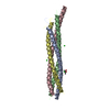 1sfcS S: Starting model for refinement |
|---|---|
| Similar structure data |


-Links
 PDBj
PDBj
- Assembly
| Deposited unit | 
| ||||||||
|---|---|---|---|---|---|---|---|---|---|
| 1 |
| ||||||||
| Unit cell |
| ||||||||
| Details | The asymmetric unit contains the biologically relevant structure |
-Components
-Protein , 3 types, 3 molecules ABE
| #1: Protein | Mass: 9046.266 Da / Num. of mol.: 1 Source method: isolated from a genetically manipulated source Source: (gene. exp.) Loligo pealei (longfin inshore squid) / Plasmid: pRSet / Production host:  Escherichia coli (E. coli) / Strain (production host): Bl21 pUBS / References: UniProt: P47194 Escherichia coli (E. coli) / Strain (production host): Bl21 pUBS / References: UniProt: P47194 |
|---|---|
| #2: Protein | Mass: 9981.440 Da / Num. of mol.: 1 Source method: isolated from a genetically manipulated source Source: (gene. exp.) Loligo pealei (longfin inshore squid) / Plasmid: pMal-TEV / Production host: Escherichia coli (E. coli) / Strain (production host): Bl21 pUBS / References: UniProt: O46345 |
| #5: Protein | Mass: 9251.200 Da / Num. of mol.: 1 Source method: isolated from a genetically manipulated source Source: (gene. exp.) Loligo pealei (longfin inshore squid) / Plasmid: pMal-TEV / Production host: Escherichia coli (E. coli) / Strain (production host): Bl21 pUBS / References: UniProt: Q95PA1 |
-S-SNAP25 fusion ... , 2 types, 2 molecules CD
| #3: Protein | Mass: 9566.525 Da / Num. of mol.: 1 Source method: isolated from a genetically manipulated source Source: (gene. exp.) Loligo pealei (longfin inshore squid) / Plasmid: pRSet / Production host: Escherichia coli (E. coli) / Strain (production host): Bl21 pUBS / References: UniProt: Q8T3S4 |
|---|---|
| #4: Protein | Mass: 9671.688 Da / Num. of mol.: 1 Source method: isolated from a genetically manipulated source Source: (gene. exp.) Loligo pealei (longfin inshore squid) / Plasmid: pRSet / Production host: Escherichia coli (E. coli) / Strain (production host): Bl21 pUBS / References: UniProt: Q8T3S4 |
-Non-polymers , 1 types, 23 molecules 
| #6: Water | ChemComp-HOH / WaterMass: 18.015 Da / Num. of mol.: 23 / Source method: isolated from a natural source / Formula: H2O |
|---|
-Experimental details
-Experiment
| Experiment | Method: X-RAY DIFFRACTION / Number of used crystals: 1 |
|---|
- Sample preparation
| Crystal | Density Matthews: 2.55 Å3/Da / Density % sol: 51.3 % | |||||||||||||||||||||||||||||||||||
|---|---|---|---|---|---|---|---|---|---|---|---|---|---|---|---|---|---|---|---|---|---|---|---|---|---|---|---|---|---|---|---|---|---|---|---|---|
| Crystal grow | Temperature: 293 K / Method: vapor diffusion, hanging drop / pH: 7.2 Details: 5% MPD, 7.5% PEG550-MME, 20MM beta mercaptoethanol, 20MM Tris PH 7.2, 50MM sodium chloride, VAPOR DIFFUSION, HANGING DROP, temperature 293K | |||||||||||||||||||||||||||||||||||
| Crystal grow | *PLUS Temperature: 20 ℃ | |||||||||||||||||||||||||||||||||||
| Components of the solutions | *PLUS
|
-Data collection
| Diffraction | Mean temperature: 100 K |
|---|---|
| Diffraction source | Source: SYNCHROTRON / Site: ESRF  / Beamline: ID29 / Wavelength: 0.915 Å / Beamline: ID29 / Wavelength: 0.915 Å |
| Detector | Type: ADSC QUANTUM 210 / Detector: CCD / Date: Nov 19, 2001 |
| Radiation | Monochromator: Si(111) / Protocol: SINGLE WAVELENGTH / Monochromatic (M) / Laue (L): M / Scattering type: x-ray |
| Radiation wavelength | Wavelength: 0.915 Å / Relative weight: 1 |
| Reflection | Resolution: 2.95→15 Å / Num. all: 10799 / Num. obs: 10128 / % possible obs: 93.6 % / Observed criterion σ(F): 0 / Observed criterion σ(I): 0 / Redundancy: 4.1 % / Biso Wilson estimate: 79.04 Å2 / Rmerge(I) obs: 0.086 / Net I/σ(I): 4.7 |
| Reflection shell | Resolution: 2.95→3.13 Å / Redundancy: 4 % / Rmerge(I) obs: 0.219 / Mean I/σ(I) obs: 3.1 / % possible all: 93 |
| Reflection | *PLUS Lowest resolution: 75 Å / Num. measured all: 41657 / Rmerge(I) obs: 0.086 |
| Reflection shell | *PLUS % possible obs: 93 % / Rmerge(I) obs: 0.219 |
- Processing
| Software |
| |||||||||||||||||||||||||
|---|---|---|---|---|---|---|---|---|---|---|---|---|---|---|---|---|---|---|---|---|---|---|---|---|---|---|
| Refinement | Method to determine structure: MOLECULAR REPLACEMENT Starting model: PDB ENTRY 1SFC Resolution: 2.95→15 Å / Rfactor Rfree error: 0.013 / Isotropic thermal model: RESTRAINED / Cross valid method: THROUGHOUT / σ(F): 0 / Stereochemistry target values: Engh & Huber
| |||||||||||||||||||||||||
| Solvent computation | Solvent model: FLAT MODEL / Bsol: 49.2327 Å2 / ksol: 0.26842 e/Å3 | |||||||||||||||||||||||||
| Displacement parameters | Biso mean: 81.5 Å2
| |||||||||||||||||||||||||
| Refine analyze |
| |||||||||||||||||||||||||
| Refinement step | Cycle: LAST / Resolution: 2.95→15 Å
| |||||||||||||||||||||||||
| Refine LS restraints |
| |||||||||||||||||||||||||
| LS refinement shell | Resolution: 2.95→3.13 Å / Rfactor Rfree error: 0.04 / Total num. of bins used: 6
| |||||||||||||||||||||||||
| Refinement | *PLUS Lowest resolution: 15 Å / Rfactor all: 0.3004 / Rfactor Rfree: 0.3445 / Rfactor Rwork: 0.297 | |||||||||||||||||||||||||
| Solvent computation | *PLUS | |||||||||||||||||||||||||
| Displacement parameters | *PLUS | |||||||||||||||||||||||||
| Refine LS restraints | *PLUS
| |||||||||||||||||||||||||
| LS refinement shell | *PLUS Rfactor Rfree: 0.397 / Rfactor Rwork: 0.346 |
