 Movie
Movie Controller


+ Open data

- Basic information
| Entry | Database: PDB / ID: 4gdo | ||||||
|---|---|---|---|---|---|---|---|
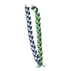
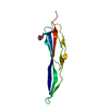
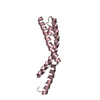
| Title | Structure of a fragment of the rod domain of plectin | ||||||
 Components Components | Plectin | ||||||
 Keywords Keywords | STRUCTURAL PROTEIN / COILED-COIL | ||||||
| Function / homology |  Function and homology information Function and homology informationprotein-containing complex organization / actomyosin contractile ring assembly actin filament organization / skeletal myofibril assembly / tight junction organization / Type I hemidesmosome assembly / hemidesmosome assembly / hemidesmosome / leukocyte migration involved in immune response / intermediate filament organization / : ...protein-containing complex organization / actomyosin contractile ring assembly actin filament organization / skeletal myofibril assembly / tight junction organization / Type I hemidesmosome assembly / hemidesmosome assembly / hemidesmosome / leukocyte migration involved in immune response / intermediate filament organization / : / regulation of vascular permeability / intermediate filament cytoskeleton organization / dystroglycan binding / cellular response to hydrostatic pressure / fibroblast migration / costamere / T cell chemotaxis / cellular response to fluid shear stress / peripheral nervous system myelin maintenance / cardiac muscle cell development / myoblast differentiation / adherens junction organization / intermediate filament cytoskeleton / response to food / structural constituent of muscle / ankyrin binding / Assembly of collagen fibrils and other multimeric structures / intermediate filament / sarcomere organization / nucleus organization / keratinocyte development / transmission of nerve impulse / brush border / sarcoplasm / Caspase-mediated cleavage of cytoskeletal proteins / establishment of skin barrier / skeletal muscle fiber development / respiratory electron transport chain / mitochondrion organization / wound healing / cell morphogenesis / protein localization / multicellular organism growth / structural constituent of cytoskeleton / sarcolemma / Z disc / cellular response to mechanical stimulus / : / actin filament binding / myelin sheath / gene expression / mitochondrial outer membrane / cadherin binding / axon / focal adhesion / dendrite / perinuclear region of cytoplasm / RNA binding / extracellular exosome / plasma membrane / cytosol / cytoplasmSimilarity search - Function | ||||||
| Biological species |  Homo sapiens (human) Homo sapiens (human) | ||||||
| Method | X-RAY DIFFRACTION / AB INITIO PHASING / Resolution: 1.7 Å | ||||||
 Authors Authors | De Pereda, J.M. / Buey, R.M. / Uson, I. / Sammito, M.D. / De Marino, I. | ||||||
 Citation Citation | Journal: Nat.Methods / Year: 2013 Title: Exploiting tertiary structure through local folds for crystallographic phasing. Authors: Sammito, M. / Millan, C. / Rodriguez, D.D. / de Ilarduya, I.M. / Meindl, K. / De Marino, I. / Petrillo, G. / Buey, R.M. / de Pereda, J.M. / Zeth, K. / Sheldrick, G.M. / Uson, I. | ||||||
| History |
|
- Structure visualization
| Structure viewer | Molecule: MolmilJmol/JSmol |
|---|
- Downloads & links
-Download
| PDBx/mmCIF format | 4gdo.cif.gz | 129 KB | Display | PDBx/mmCIF format |
|---|---|---|---|---|
| PDB format | pdb4gdo.ent.gz | 106.4 KB | Display | PDB format |
| PDBx/mmJSON format | 4gdo.json.gz | Tree view | PDBx/mmJSON format | |
| Others |  Other downloads Other downloads |
-Validation report
| Arichive directory | https://data.pdbj.org/pub/pdb/validation_reports/gd/4gdoftp://data.pdbj.org/pub/pdb/validation_reports/gd/4gdo | HTTPS FTP |
|---|
-Related structure data


-Links
 PDBj
PDBj


- Assembly
| Deposited unit | 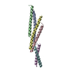
| ||||||||
|---|---|---|---|---|---|---|---|---|---|
| 1 | 
| ||||||||
| 2 | 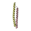
| ||||||||
| 3 | 
| ||||||||
| Unit cell |
| ||||||||
| Details | AS PER THE AUTHORS THE BIOLOGICAL ASSEMBLY IS UNKNOWN |
-Components
| #1: Protein/peptide | / PCN / PLTN / Hemidesmosomal protein 1 / HD1 / Plectin-1 Mass: 4880.413 Da / Num. of mol.: 6 Fragment: FRAGMENT OF THE ROD DOMAIN, UNP residues 1492-1530 Source method: isolated from a genetically manipulated source Source: (gene. exp.) Homo sapiens (human) / Gene: PLEC, PLEC1 / Plasmid: Modified pGEX-4T3 / Production host:  Escherichia coli (E. coli) / Strain (production host): BL21(DE3)T1 / References: UniProt: Q15149 Escherichia coli (E. coli) / Strain (production host): BL21(DE3)T1 / References: UniProt: Q15149#2: Water | ChemComp-HOH / | Water Mass: 18.015 Da / Num. of mol.: 163 / Source method: isolated from a natural source / Formula: H2O Mass: 18.015 Da / Num. of mol.: 163 / Source method: isolated from a natural source / Formula: H2O |
|---|
-Experimental details
-Experiment
| Experiment | Method: X-RAY DIFFRACTION / Number of used crystals: 1 |
|---|
- Sample preparation
| Crystal | Density Matthews: 2.1 Å3/Da / Density % sol: 41.29 % |
|---|---|
| Crystal grow | Temperature: 298 K / Method: vapor diffusion, sitting drop / pH: 4.5 Details: 0.1M sodium acetate, 2.2M sodium chloride, 0.2M lithium sulfate, pH 4.5, VAPOR DIFFUSION, SITTING DROP, temperature 298K |
-Data collection
| Diffraction | Mean temperature: 100 K |
|---|---|
| Diffraction source | Source: ROTATING ANODE / Type: BRUKER AXS MICROSTAR-H / Wavelength: 1.5418 Å |
| Detector | Type: MAR scanner 345 mm plate / Detector: IMAGE PLATE / Date: Apr 1, 2012 |
| Radiation | Monochromator: HELIOS OPTICS / Protocol: SINGLE WAVELENGTH / Monochromatic (M) / Laue (L): M / Scattering type: x-ray |
| Radiation wavelength | Wavelength: 1.5418 Å / Relative weight: 1 |
| Reflection | Resolution: 1.7→77 Å / Num. all: 23087 / Num. obs: 23087 / % possible obs: 85.3 % / Observed criterion σ(F): 0 / Observed criterion σ(I): -3 / Redundancy: 3.1 % |
| Reflection shell | Resolution: 1.7→1.74 Å / Redundancy: 2.1 % / Mean I/σ(I) obs: 3.52 / Num. unique all: 1381 / % possible all: 68.9 |
- Processing
| Software |
| |||||||||||||||||||||||||||||||||||||||||||||||||||||||||||||||||||||||||||||||||||||||||||||||||||||||||||||||||||||||||||||||||||||||||||||||||||||||||||||||||||||||||||||||
|---|---|---|---|---|---|---|---|---|---|---|---|---|---|---|---|---|---|---|---|---|---|---|---|---|---|---|---|---|---|---|---|---|---|---|---|---|---|---|---|---|---|---|---|---|---|---|---|---|---|---|---|---|---|---|---|---|---|---|---|---|---|---|---|---|---|---|---|---|---|---|---|---|---|---|---|---|---|---|---|---|---|---|---|---|---|---|---|---|---|---|---|---|---|---|---|---|---|---|---|---|---|---|---|---|---|---|---|---|---|---|---|---|---|---|---|---|---|---|---|---|---|---|---|---|---|---|---|---|---|---|---|---|---|---|---|---|---|---|---|---|---|---|---|---|---|---|---|---|---|---|---|---|---|---|---|---|---|---|---|---|---|---|---|---|---|---|---|---|---|---|---|---|---|---|---|---|
| Refinement | Method to determine structure: AB INITIO PHASING / Resolution: 1.7→45.194 Å / SU ML: 0.19 / σ(F): 1.68 / Phase error: 27.63 / Stereochemistry target values: ML
| |||||||||||||||||||||||||||||||||||||||||||||||||||||||||||||||||||||||||||||||||||||||||||||||||||||||||||||||||||||||||||||||||||||||||||||||||||||||||||||||||||||||||||||||
| Solvent computation | Shrinkage radii: 0.9 Å / VDW probe radii: 1.11 Å / Solvent model: FLAT BULK SOLVENT MODEL | |||||||||||||||||||||||||||||||||||||||||||||||||||||||||||||||||||||||||||||||||||||||||||||||||||||||||||||||||||||||||||||||||||||||||||||||||||||||||||||||||||||||||||||||
| Refinement step | Cycle: LAST / Resolution: 1.7→45.194 Å
| |||||||||||||||||||||||||||||||||||||||||||||||||||||||||||||||||||||||||||||||||||||||||||||||||||||||||||||||||||||||||||||||||||||||||||||||||||||||||||||||||||||||||||||||
| Refine LS restraints |
| |||||||||||||||||||||||||||||||||||||||||||||||||||||||||||||||||||||||||||||||||||||||||||||||||||||||||||||||||||||||||||||||||||||||||||||||||||||||||||||||||||||||||||||||
| LS refinement shell |
| |||||||||||||||||||||||||||||||||||||||||||||||||||||||||||||||||||||||||||||||||||||||||||||||||||||||||||||||||||||||||||||||||||||||||||||||||||||||||||||||||||||||||||||||
| Refinement TLS params. | Method: refined / Refine-ID: X-RAY DIFFRACTION
| |||||||||||||||||||||||||||||||||||||||||||||||||||||||||||||||||||||||||||||||||||||||||||||||||||||||||||||||||||||||||||||||||||||||||||||||||||||||||||||||||||||||||||||||
| Refinement TLS group |
|
