 Movie
Movie Controller

[English] 日本語
 Yorodumi
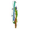
- PDB-2d42: Crystal structure analysis of a non-toxic crystal protein from Ba... -
+ Open data

- Basic information
| Entry | Database: PDB / ID: 2d42 | ||||||
|---|---|---|---|---|---|---|---|
| Title | Crystal structure analysis of a non-toxic crystal protein from Bacillus thuringiensis | ||||||
 Components Components | non-toxic crystal protein | ||||||
 Keywords Keywords |  TOXIN / parasporin / bacterial toxin / beta-pore-forming toxin / parasporal inclusion / hinge-bending motion TOXIN / parasporin / bacterial toxin / beta-pore-forming toxin / parasporal inclusion / hinge-bending motion | ||||||
| Function / homology |  Function and homology information Function and homology informationNuclear Transport Factor 2; Chain: A, - #380 / Proaerolysin, chain A, domain 3 / Aerolysin-like toxin / Clostridium epsilon toxin ETX/Bacillus mosquitocidal toxin MTX2 / Proaerolysin; Chain A, domain 3 / Nuclear Transport Factor 2; Chain: A, / Beta Complex / Roll / Mainly Beta / Alpha Beta Similarity search - Domain/homology | ||||||
| Biological species |  Bacillus thuringiensis (bacteria) Bacillus thuringiensis (bacteria) | ||||||
| Method | X-RAY DIFFRACTION / SYNCHROTRON / MAD / Resolution: 2.07 Å | ||||||
 Authors Authors | Akiba, T. / Higuchi, K. / Mizuki, E. / Ekino, K. / Shin, T. / Ohba, M. / Kanai, R. / Harata, K. | ||||||
 Citation Citation | Journal: Proteins / Year: 2006 Title: Nontoxic crystal protein from Bacillus thuringiensis demonstrates a remarkable structural similarity to beta-pore-forming toxins Authors: Akiba, T. / Higuchi, K. / Mizuki, E. / Ekino, K. / Shin, T. / Ohba, M. / Kanai, R. / Harata, K. | ||||||
| History |
|
- Structure visualization
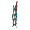
| Structure viewer | Molecule: MolmilJmol/JSmol |
|---|
- Downloads & links
-Download
| PDBx/mmCIF format | 2d42.cif.gz | 109.4 KB | Display | PDBx/mmCIF format |
|---|---|---|---|---|
| PDB format | pdb2d42.ent.gz | 86 KB | Display | PDB format |
| PDBx/mmJSON format | 2d42.json.gz | Tree view | PDBx/mmJSON format | |
| Others |  Other downloads Other downloads |
-Validation report
| Arichive directory | https://data.pdbj.org/pub/pdb/validation_reports/d4/2d42ftp://data.pdbj.org/pub/pdb/validation_reports/d4/2d42 | HTTPS FTP |
|---|
-Related structure data
| Similar structure data |
|---|


-Links
 PDBj
PDBj- Assembly
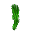
| Deposited unit | 
| ||||||||
|---|---|---|---|---|---|---|---|---|---|
| 1 | 
| ||||||||
| 2 | 
| ||||||||
| Unit cell |
|
-Components
| #1: Protein | Mass: 27565.605 Da / Num. of mol.: 2 / Source method: isolated from a natural source / Source: (natural) Bacillus thuringiensis (bacteria) / Strain: A1470 (formerly called 89-T-34-22) / References: UniProt: Q6L5X8*PLUS#2: Water | ChemComp-HOH / | Water Mass: 18.015 Da / Num. of mol.: 210 / Source method: isolated from a natural source / Formula: H2O Mass: 18.015 Da / Num. of mol.: 210 / Source method: isolated from a natural source / Formula: H2O |
|---|
-Experimental details
-Experiment
| Experiment | Method: X-RAY DIFFRACTION / Number of used crystals: 2 |
|---|
- Sample preparation
| Crystal | Density Matthews: 3.1 Å3/Da / Density % sol: 60.3 % |
|---|---|
| Crystal grow | Temperature: 298 K / Method: vapor diffusion, hanging drop / pH: 5.8 Details: 10%(v/v) 2-methyl-2,4-pentanediol, 50-100mM ammonium sulfate, 100mM sodium citrate, 1mM Na-EDTA, 1mM DTT, pH 5.8, VAPOR DIFFUSION, HANGING DROP, temperature 298K |
-Data collection
| Diffraction |
| ||||||||||||||||||
|---|---|---|---|---|---|---|---|---|---|---|---|---|---|---|---|---|---|---|---|
| Diffraction source |
| ||||||||||||||||||
| Detector |
| ||||||||||||||||||
| Radiation |
| ||||||||||||||||||
| Radiation wavelength |
| ||||||||||||||||||
| Reflection | Resolution: 2.07→47.67 Å / Num. obs: 41642 / % possible obs: 99.9 % / Redundancy: 11.9 % / Biso Wilson estimate: 20.4 Å2 / Rsym value: 0.054 / Net I/σ(I): 32.8 | ||||||||||||||||||
| Reflection shell | Resolution: 2.07→2.18 Å / Redundancy: 10.9 % / Mean I/σ(I) obs: 9.6 / Num. unique all: 6001 / Rsym value: 0.229 / % possible all: 99.8 |

- Processing
| Software |
| ||||||||||||||||||||||||||||||||||||||||||||||||||||||||||||||||||||||||||||||||
|---|---|---|---|---|---|---|---|---|---|---|---|---|---|---|---|---|---|---|---|---|---|---|---|---|---|---|---|---|---|---|---|---|---|---|---|---|---|---|---|---|---|---|---|---|---|---|---|---|---|---|---|---|---|---|---|---|---|---|---|---|---|---|---|---|---|---|---|---|---|---|---|---|---|---|---|---|---|---|---|---|---|
| Refinement | Method to determine structure: MAD / Resolution: 2.07→47.67 Å / Rfactor Rfree error: 0.004 / Data cutoff high absF: 2145829.23 / Data cutoff low absF: 0 / Isotropic thermal model: RESTRAINED / Cross valid method: THROUGHOUT / σ(F): 0 / Stereochemistry target values: Engh & Huber
| ||||||||||||||||||||||||||||||||||||||||||||||||||||||||||||||||||||||||||||||||
| Solvent computation | Solvent model: FLAT MODEL / Bsol: 49.4809 Å2 / ksol: 0.380301 e/Å3 | ||||||||||||||||||||||||||||||||||||||||||||||||||||||||||||||||||||||||||||||||
| Displacement parameters | Biso mean: 35.5 Å2
| ||||||||||||||||||||||||||||||||||||||||||||||||||||||||||||||||||||||||||||||||
| Refine analyze |
| ||||||||||||||||||||||||||||||||||||||||||||||||||||||||||||||||||||||||||||||||
| Refinement step | Cycle: LAST / Resolution: 2.07→47.67 Å
| ||||||||||||||||||||||||||||||||||||||||||||||||||||||||||||||||||||||||||||||||
| Refine LS restraints |
| ||||||||||||||||||||||||||||||||||||||||||||||||||||||||||||||||||||||||||||||||
| LS refinement shell | Resolution: 2.07→2.2 Å / Rfactor Rfree error: 0.011 / Total num. of bins used: 6
| ||||||||||||||||||||||||||||||||||||||||||||||||||||||||||||||||||||||||||||||||
| Xplor file |
|
