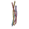
 Movie
Movie Controller


+ Open data

- Basic information
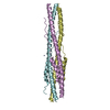
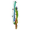
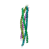
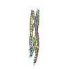
| Entry | Database: PDB / ID: 4wy4 | |||||||||
|---|---|---|---|---|---|---|---|---|---|---|
| Title | Crystal structure of autophagic SNARE complex | |||||||||
 Components Components |
| |||||||||
 Keywords Keywords |  MEMBRANE PROTEIN / autophagy / SNARE / fusion MEMBRANE PROTEIN / autophagy / SNARE / fusion | |||||||||
| Function / homology |  Function and homology information Function and homology informationendoplasmic reticulum-Golgi intermediate compartment organization / mucin granule / negative regulation of secretion by cell / ciliary pocket membrane / positive regulation of histamine secretion by mast cell / smooth endoplasmic reticulum membrane / vesicle targeting / trans-Golgi Network Vesicle Budding / synaptic vesicle fusion to presynaptic active zone membrane / mitochondria-associated endoplasmic reticulum membrane contact site ...endoplasmic reticulum-Golgi intermediate compartment organization / mucin granule / negative regulation of secretion by cell / ciliary pocket membrane / positive regulation of histamine secretion by mast cell / smooth endoplasmic reticulum membrane / vesicle targeting / trans-Golgi Network Vesicle Budding / synaptic vesicle fusion to presynaptic active zone membrane / mitochondria-associated endoplasmic reticulum membrane contact site / Intra-Golgi traffic / mucus secretion / vesicle fusion / vesicle docking / chloride channel inhibitor activity / SNARE complex / SNAP receptor activity / protein localization to phagophore assembly site / SNARE complex assembly / COPII-mediated vesicle transport / synaptic vesicle priming / syntaxin binding / Lysosome Vesicle Biogenesis / azurophil granule membrane / autophagosome membrane docking / Golgi Associated Vesicle Biogenesis / COPII-coated ER to Golgi transport vesicle / Golgi organization / exocytosis / autophagosome maturation / autophagosome membrane / tertiary granule membrane / cilium assembly / endoplasmic reticulum-Golgi intermediate compartment / autophagosome / endoplasmic reticulum to Golgi vesicle-mediated transport / endomembrane system / specific granule membrane / regulation of protein localization to plasma membrane / endoplasmic reticulum-Golgi intermediate compartment membrane / SNARE binding / secretory granule membrane / intracellular protein transport / clathrin-coated endocytic vesicle membrane / ER to Golgi transport vesicle membrane / recycling endosome / recycling endosome membrane / phagocytic vesicle membrane / protein transport / Cargo recognition for clathrin-mediated endocytosis / presynapse / Clathrin-mediated endocytosis / late endosome membrane / ER-Phagosome pathway / early endosome membrane / protein phosphatase binding / defense response to virus / vesicle / membrane fusion / early endosome / symbiont entry into host cell / lysosomal membrane / Golgi membrane / centrosome / Neutrophil degranulation / endoplasmic reticulum membrane / protein kinase binding / perinuclear region of cytoplasm / mitochondrion / extracellular exosome / membrane / plasma membrane / cytosol / cytoplasmSimilarity search - Function | |||||||||
| Biological species |  Homo sapiens (human) Homo sapiens (human) | |||||||||
| Method | X-RAY DIFFRACTION / SYNCHROTRON / Resolution: 1.4 Å | |||||||||
 Authors Authors | Zhao, M. / Brunger, A.T. | |||||||||
| Funding support |  United States, 2items United States, 2items
| |||||||||
 Citation Citation | Journal: Nature / Year: 2015 Title: ATG14 promotes membrane tethering and fusion of autophagosomes to endolysosomes. Authors: Diao, J. / Liu, R. / Rong, Y. / Zhao, M. / Zhang, J. / Lai, Y. / Zhou, Q. / Wilz, L.M. / Li, J. / Vivona, S. / Pfuetzner, R.A. / Brunger, A.T. / Zhong, Q. | |||||||||
| History |
|
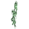
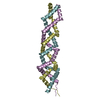
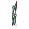
- Structure visualization
| Structure viewer | Molecule: MolmilJmol/JSmol |
|---|
- Downloads & links
-Download
| PDBx/mmCIF format | 4wy4.cif.gz | 139.2 KB | Display | PDBx/mmCIF format |
|---|---|---|---|---|
| PDB format | pdb4wy4.ent.gz | 109.6 KB | Display | PDB format |
| PDBx/mmJSON format | 4wy4.json.gz | Tree view | PDBx/mmJSON format | |
| Others |  Other downloads Other downloads |
-Validation report
| Arichive directory | https://data.pdbj.org/pub/pdb/validation_reports/wy/4wy4ftp://data.pdbj.org/pub/pdb/validation_reports/wy/4wy4 | HTTPS FTP |
|---|
-Related structure data
| Similar structure data |
|---|


-Links
 PDBj
PDBj


- Assembly
| Deposited unit | 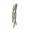
| ||||||||
|---|---|---|---|---|---|---|---|---|---|
| 1 |
| ||||||||
| Unit cell |
| ||||||||
| Details | biological unit is the same as asym. |
-Components
| #1: Protein | / VAMP-8 / Endobrevin / EDB Mass: 7623.581 Da / Num. of mol.: 1 Source method: isolated from a genetically manipulated source Source: (gene. exp.) Homo sapiens (human) / Gene: VAMP8 / Plasmid: pACYCDuet-1 / Production host:  Escherichia coli (E. coli) / Strain (production host): BL21(DE3) / References: UniProt: Q9BV40 Escherichia coli (E. coli) / Strain (production host): BL21(DE3) / References: UniProt: Q9BV40 |
|---|---|
| #2: Protein | Mass: 6367.963 Da / Num. of mol.: 1 Source method: isolated from a genetically manipulated source Source: (gene. exp.) Homo sapiens (human) / Gene: STX17 / Plasmid: pACYCDuet-1 / Production host: Escherichia coli (E. coli) / Strain (production host): BL21(DE3) / References: UniProt: P56962 |
| #3: Protein | Mass: 8928.136 Da / Num. of mol.: 1 Source method: isolated from a genetically manipulated source Source: (gene. exp.) Homo sapiens (human) / Gene: SNAP29 / Plasmid: pETDuet-1 / Production host: Escherichia coli (E. coli) / Strain (production host): BL21(DE3) / References: UniProt: O95721 |
| #4: Protein | Mass: 7561.632 Da / Num. of mol.: 1 Source method: isolated from a genetically manipulated source Source: (gene. exp.) Homo sapiens (human) / Gene: SNAP29 / Plasmid: pETDuet-1 / Production host: Escherichia coli (E. coli) / Strain (production host): BL21(DE3) / References: UniProt: O95721 |
| #5: Water | ChemComp-HOH / Water Mass: 18.015 Da / Num. of mol.: 299 / Source method: isolated from a natural source / Formula: H2O Mass: 18.015 Da / Num. of mol.: 299 / Source method: isolated from a natural source / Formula: H2O |
-Experimental details
-Experiment
| Experiment | Method: X-RAY DIFFRACTION / Number of used crystals: 1 |
|---|
- Sample preparation
| Crystal | Density Matthews: 2.47 Å3/Da / Density % sol: 50.28 % |
|---|---|
| Crystal grow | Temperature: 295 K / Method: vapor diffusion, hanging drop / pH: 8.5 / Details: 0.1M BICINE pH 8.5, 55% (v/v) MPD |
-Data collection
| Diffraction | Mean temperature: 100 K | ||||||||||||||||||||||||||||||||||||||||||||||||||||||||||||||||||||||||||||||||||||||||||||||||||||||||||||||||||||||||||||||||||||||||||||||||||||||||||||||||||||||||||||||||||||||||||||||||||||||||||||||||||
|---|---|---|---|---|---|---|---|---|---|---|---|---|---|---|---|---|---|---|---|---|---|---|---|---|---|---|---|---|---|---|---|---|---|---|---|---|---|---|---|---|---|---|---|---|---|---|---|---|---|---|---|---|---|---|---|---|---|---|---|---|---|---|---|---|---|---|---|---|---|---|---|---|---|---|---|---|---|---|---|---|---|---|---|---|---|---|---|---|---|---|---|---|---|---|---|---|---|---|---|---|---|---|---|---|---|---|---|---|---|---|---|---|---|---|---|---|---|---|---|---|---|---|---|---|---|---|---|---|---|---|---|---|---|---|---|---|---|---|---|---|---|---|---|---|---|---|---|---|---|---|---|---|---|---|---|---|---|---|---|---|---|---|---|---|---|---|---|---|---|---|---|---|---|---|---|---|---|---|---|---|---|---|---|---|---|---|---|---|---|---|---|---|---|---|---|---|---|---|---|---|---|---|---|---|---|---|---|---|---|---|---|
| Diffraction source | Source: SYNCHROTRON / Site: APS / Beamline: 24-ID-C / Wavelength: 0.9792 Å | ||||||||||||||||||||||||||||||||||||||||||||||||||||||||||||||||||||||||||||||||||||||||||||||||||||||||||||||||||||||||||||||||||||||||||||||||||||||||||||||||||||||||||||||||||||||||||||||||||||||||||||||||||
| Detector | Type: PSI PILATUS 6M / Detector: PIXEL / Date: Nov 15, 2013 | ||||||||||||||||||||||||||||||||||||||||||||||||||||||||||||||||||||||||||||||||||||||||||||||||||||||||||||||||||||||||||||||||||||||||||||||||||||||||||||||||||||||||||||||||||||||||||||||||||||||||||||||||||
| Radiation | Protocol: SINGLE WAVELENGTH / Monochromatic (M) / Laue (L): M / Scattering type: x-ray | ||||||||||||||||||||||||||||||||||||||||||||||||||||||||||||||||||||||||||||||||||||||||||||||||||||||||||||||||||||||||||||||||||||||||||||||||||||||||||||||||||||||||||||||||||||||||||||||||||||||||||||||||||
| Radiation wavelength | Wavelength: 0.9792 Å / Relative weight: 1 | ||||||||||||||||||||||||||||||||||||||||||||||||||||||||||||||||||||||||||||||||||||||||||||||||||||||||||||||||||||||||||||||||||||||||||||||||||||||||||||||||||||||||||||||||||||||||||||||||||||||||||||||||||
| Reflection | Resolution: 1.4→51.97 Å / Num. obs: 58501 / % possible obs: 98.7 % / Observed criterion σ(I): -3 / Redundancy: 6.6 % / Biso Wilson estimate: 17.3 Å2 / Rmerge F obs: 0.997 / Rmerge(I) obs: 0.063 / Rrim(I) all: 0.068 / Χ2: 1.026 / Net I/σ(I): 14.32 / Num. measured all: 385970 | ||||||||||||||||||||||||||||||||||||||||||||||||||||||||||||||||||||||||||||||||||||||||||||||||||||||||||||||||||||||||||||||||||||||||||||||||||||||||||||||||||||||||||||||||||||||||||||||||||||||||||||||||||
| Reflection shell | Diffraction-ID: 1 / Rejects: 0
|
- Processing
| Software |
| ||||||||||||||||||||||||||||||||||||||||||||||||||||||||||||||||||||||||||||||||||||||||||||||||||||||||||||||||||||||||||||||||||||||||||||||||||||||||||
|---|---|---|---|---|---|---|---|---|---|---|---|---|---|---|---|---|---|---|---|---|---|---|---|---|---|---|---|---|---|---|---|---|---|---|---|---|---|---|---|---|---|---|---|---|---|---|---|---|---|---|---|---|---|---|---|---|---|---|---|---|---|---|---|---|---|---|---|---|---|---|---|---|---|---|---|---|---|---|---|---|---|---|---|---|---|---|---|---|---|---|---|---|---|---|---|---|---|---|---|---|---|---|---|---|---|---|---|---|---|---|---|---|---|---|---|---|---|---|---|---|---|---|---|---|---|---|---|---|---|---|---|---|---|---|---|---|---|---|---|---|---|---|---|---|---|---|---|---|---|---|---|---|---|---|---|
| Refinement | Resolution: 1.4→51.967 Å / SU ML: 0.13 / Cross valid method: FREE R-VALUE / σ(F): 1.36 / Phase error: 16.95 / Stereochemistry target values: ML
| ||||||||||||||||||||||||||||||||||||||||||||||||||||||||||||||||||||||||||||||||||||||||||||||||||||||||||||||||||||||||||||||||||||||||||||||||||||||||||
| Solvent computation | Shrinkage radii: 0.9 Å / VDW probe radii: 1.11 Å / Solvent model: FLAT BULK SOLVENT MODEL | ||||||||||||||||||||||||||||||||||||||||||||||||||||||||||||||||||||||||||||||||||||||||||||||||||||||||||||||||||||||||||||||||||||||||||||||||||||||||||
| Displacement parameters | Biso max: 81.53 Å2 / Biso mean: 28.9173 Å2 / Biso min: 11.88 Å2 | ||||||||||||||||||||||||||||||||||||||||||||||||||||||||||||||||||||||||||||||||||||||||||||||||||||||||||||||||||||||||||||||||||||||||||||||||||||||||||
| Refinement step | Cycle: final / Resolution: 1.4→51.967 Å
| ||||||||||||||||||||||||||||||||||||||||||||||||||||||||||||||||||||||||||||||||||||||||||||||||||||||||||||||||||||||||||||||||||||||||||||||||||||||||||
| Refine LS restraints |
| ||||||||||||||||||||||||||||||||||||||||||||||||||||||||||||||||||||||||||||||||||||||||||||||||||||||||||||||||||||||||||||||||||||||||||||||||||||||||||
| LS refinement shell | Refine-ID: X-RAY DIFFRACTION / Total num. of bins used: 21
|
