 Movie
Movie Controller


+ Open data

- Basic information
| Entry | Database: PDB / ID: 2nps | ||||||
|---|---|---|---|---|---|---|---|
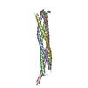
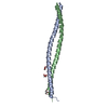
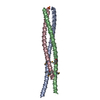
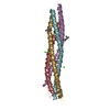
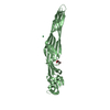
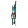
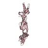
| Title | Crystal Structure of the Early Endosomal SNARE Complex | ||||||
 Components Components |
| ||||||
 Keywords Keywords |  TRANSPORT PROTEIN / Vesicle fusion / SNARE complex / early endosomal SNARE complex / syntaxin 6 / syntaxin 13 / Vti1a / VAMP4 TRANSPORT PROTEIN / Vesicle fusion / SNARE complex / early endosomal SNARE complex / syntaxin 6 / syntaxin 13 / Vti1a / VAMP4 | ||||||
| Function / homology |  Function and homology information Function and homology informationregulation of Golgi to plasma membrane protein transport / : / synaptic vesicle to endosome fusion / Retrograde transport at the Trans-Golgi-Network / : / Intra-Golgi traffic / Retrograde transport at the Trans-Golgi-Network / postsynaptic recycling endosome / vesicle fusion with Golgi apparatus / Retrograde transport at the Trans-Golgi-Network ...regulation of Golgi to plasma membrane protein transport / : / synaptic vesicle to endosome fusion / Retrograde transport at the Trans-Golgi-Network / : / Intra-Golgi traffic / Retrograde transport at the Trans-Golgi-Network / postsynaptic recycling endosome / vesicle fusion with Golgi apparatus / Retrograde transport at the Trans-Golgi-Network / : / Golgi ribbon formation / Intra-Golgi traffic / Golgi vesicle transport / Golgi to vacuole transport / vesicle fusion / vesicle docking / clathrin-coated vesicle membrane / voluntary musculoskeletal movement / SNARE complex / Golgi to plasma membrane protein transport / Cargo recognition for clathrin-mediated endocytosis / SNAP receptor activity / intra-Golgi vesicle-mediated transport / protein targeting to vacuole / Clathrin-mediated endocytosis / endocytic recycling / neuron projection terminus / phagophore assembly site / retrograde transport, endosome to Golgi / SNARE complex assembly / syntaxin binding / cholesterol efflux / clathrin-coated vesicle / regulation of synaptic vesicle endocytosis / autophagosome assembly / autophagosome / endomembrane system / endoplasmic reticulum to Golgi vesicle-mediated transport / phagocytic vesicle / SNARE binding / trans-Golgi network membrane / intracellular protein transport / trans-Golgi network / ER to Golgi transport vesicle membrane / terminal bouton / synaptic vesicle membrane / microtubule cytoskeleton organization / recycling endosome / autophagy / cellular response to type II interferon / recycling endosome membrane / regulation of protein localization / synaptic vesicle / late endosome membrane / early endosome membrane / vesicle / membrane => GO:0016020 / protein stabilization / early endosome / endosome / membrane raft / Golgi membrane / neuronal cell body / endoplasmic reticulum membrane / perinuclear region of cytoplasm / Golgi apparatus / cell surface / nucleoplasm / plasma membrane / cytosolSimilarity search - Function | ||||||
| Biological species |  Mus musculus (house mouse)Rattus norvegicus (Norway rat) Mus musculus (house mouse)Rattus norvegicus (Norway rat) Homo sapiens (human) Homo sapiens (human) | ||||||
| Method | X-RAY DIFFRACTION / SYNCHROTRON / MOLECULAR REPLACEMENT / Resolution: 2.5 Å | ||||||
 Authors Authors | Zwilling, D. / Cypionka, A. / Pohl, W.H. / Fasshauer, D. / Walla, P.J. / Wahl, M.C. / Jahn, R. | ||||||
 Citation Citation | Journal: Embo J. / Year: 2007 Title: Early endosomal SNAREs form a structurally conserved SNARE complex and fuse liposomes with multiple topologies Authors: Zwilling, D. / Cypionka, A. / Pohl, W.H. / Fasshauer, D. / Walla, P.J. / Wahl, M.C. / Jahn, R. | ||||||
| History |
|
- Structure visualization
| Structure viewer | Molecule: MolmilJmol/JSmol |
|---|
- Downloads & links
-Download
| PDBx/mmCIF format | 2nps.cif.gz | 72.1 KB | Display | PDBx/mmCIF format |
|---|---|---|---|---|
| PDB format | pdb2nps.ent.gz | 52.7 KB | Display | PDB format |
| PDBx/mmJSON format | 2nps.json.gz | Tree view | PDBx/mmJSON format | |
| Others |  Other downloads Other downloads |
-Validation report
| Arichive directory | https://data.pdbj.org/pub/pdb/validation_reports/np/2npsftp://data.pdbj.org/pub/pdb/validation_reports/np/2nps | HTTPS FTP |
|---|
-Related structure data
| Related structure data | 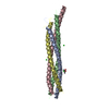 1sfcS S: Starting model for refinement |
|---|---|
| Similar structure data |

-Links
 PDBj
PDBj


- Assembly
| Deposited unit | 
| ||||||||
|---|---|---|---|---|---|---|---|---|---|
| 1 |
| ||||||||
| Unit cell |
| ||||||||
| Details | The biological assembly is a tetrameric coiled-coil comprised of chains A, B, C, and D. There is one biological assembly per asymmetric unit. |
-Components
| #1: Protein | Mass: 8687.821 Da / Num. of mol.: 1 / Fragment: VAMP4 SNARE Motif, residues 47-117 Source method: isolated from a genetically manipulated source Source: (gene. exp.) Mus musculus (house mouse) / Gene: Vamp4_predicted / Plasmid: pET28a / Species (production host): Escherichia coli / Production host:  Escherichia coli BL21(DE3) (bacteria) / Strain (production host): BL21(DE3) / References: UniProt: O70480 Escherichia coli BL21(DE3) (bacteria) / Strain (production host): BL21(DE3) / References: UniProt: O70480 |
|---|---|
| #2: Protein | Mass: 8149.168 Da / Num. of mol.: 1 / Fragment: Syntaxin 13 SNARE Motif, residues 177-244 Source method: isolated from a genetically manipulated source Source: (gene. exp.) Rattus norvegicus (Norway rat) / Gene: syntaxin 13 / Plasmid: pEt28a / Species (production host): Escherichia coli / Production host: Escherichia coli BL21(DE3) (bacteria) / Strain (production host): BL21(DE3) / References: UniProt: O70319, UniProt: G3V7P1*PLUS |
| #3: Protein | Mass: 9548.797 Da / Num. of mol.: 1 / Fragment: Vti1a SNARE Motif, residues 122-199 Source method: isolated from a genetically manipulated source Source: (gene. exp.) Rattus norvegicus (Norway rat) / Gene: Vti1a / Plasmid: pET28a / Species (production host): Escherichia coli / Production host: Escherichia coli BL21(DE3) (bacteria) / Strain (production host): BL21(DE3) / References: UniProt: Q9JI51 |
| #4: Protein | Mass: 9121.297 Da / Num. of mol.: 1 Fragment: Syntaxin 6 SNARE Motif, t-SNARE coiled-coil homology, residues 169-234 Source method: isolated from a genetically manipulated source Source: (gene. exp.) Homo sapiens (human) / Gene: STX6 / Plasmid: pET28a / Species (production host): Escherichia coli / Production host: Escherichia coli BL21(DE3) (bacteria) / Strain (production host): BL21(DE3) / References: UniProt: O43752 |
| #5: Water | ChemComp-HOH / Water Mass: 18.015 Da / Num. of mol.: 121 / Source method: isolated from a natural source / Formula: H2O Mass: 18.015 Da / Num. of mol.: 121 / Source method: isolated from a natural source / Formula: H2O |
-Experimental details
-Experiment
| Experiment | Method: X-RAY DIFFRACTION / Number of used crystals: 5 |
|---|
- Sample preparation
| Crystal | Density Matthews: 2.12 Å3/Da / Density % sol: 41.85 % |
|---|---|
| Crystal grow | Temperature: 293 K / Method: vapor diffusion, sitting drop / pH: 5.6 Details: 0.1M trisodium citrate dihydrate, 36% (v/v) 2-methyl-2,4-pentandiol, 0.2M Li2SO4, pH 5.6, VAPOR DIFFUSION, SITTING DROP, temperature 293K |
-Data collection
| Diffraction | Mean temperature: 100 K |
|---|---|
| Diffraction source | Source: SYNCHROTRON / Site: SLS  / Beamline: X10SA / Wavelength: 0.9 Å / Beamline: X10SA / Wavelength: 0.9 Å |
| Detector | Type: MAR CCD 165 mm / Detector: CCD / Date: Feb 25, 2005 / Details: mirrors |
| Radiation | Protocol: SINGLE WAVELENGTH / Monochromatic (M) / Laue (L): M / Scattering type: x-ray |
| Radiation wavelength | Wavelength: 0.9 Å / Relative weight: 1 |
| Reflection | Resolution: 2.5→30 Å / Num. all: 10740 / Num. obs: 9365 / % possible obs: 87.2 % / Observed criterion σ(F): 0 / Observed criterion σ(I): 0 / Redundancy: 5.3 % / Biso Wilson estimate: 57.4 Å2 / Rmerge(I) obs: 0.124 / Rsym value: 0.039 / Net I/σ(I): 8.7 |
| Reflection shell | Resolution: 2.5→2.66 Å / Redundancy: 5.1 % / Rmerge(I) obs: 0.49 / Mean I/σ(I) obs: 1.9 / Num. unique all: 827 / Rsym value: 0.309 / % possible all: 46.4 |
- Processing
| Software |
| |||||||||||||||||||||||||
|---|---|---|---|---|---|---|---|---|---|---|---|---|---|---|---|---|---|---|---|---|---|---|---|---|---|---|
| Refinement | Method to determine structure: MOLECULAR REPLACEMENT Starting model: PDB Entry 1SFC Resolution: 2.5→30 Å / Isotropic thermal model: isotropic / Cross valid method: THROUGHOUT / σ(F): 0 / σ(I): 0 / Stereochemistry target values: Engh & Huber
| |||||||||||||||||||||||||
| Displacement parameters | Biso mean: 53.5 Å2 | |||||||||||||||||||||||||
| Refinement step | Cycle: LAST / Resolution: 2.5→30 Å
| |||||||||||||||||||||||||
| Refine LS restraints |
| |||||||||||||||||||||||||
| LS refinement shell | Resolution: 2.5→2.564 Å / Rfactor Rfree error: 0.299
|
