Mass: 45353.691 Da / Num. of mol.: 1 / Mutation: YES Source method: isolated from a genetically manipulated source Source: (gene. exp.) HOMO SAPIENS (human) / Tissue: BLOOD PLASMA / Description: THE MUTATION IS A NATURALLY OCCURRING VARIANT / Plasmid: PZM-S / Cellular location (production host): CYTOPLASM / Production host: ESCHERICHIA COLI (E. coli) / Strain (production host): N4830-1 / References: UniProt: P01011
Compound details
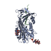
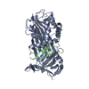
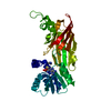
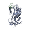
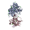
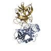
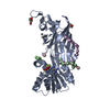
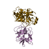
RESIDUE 55 HAS BEEN MUTATED FROM LEU TO PRO. THIS MUTANT IS A NATURALLY OCCURRING VARIANT WHICH HAS ...RESIDUE 55 HAS BEEN MUTATED FROM LEU TO PRO. THIS MUTANT IS A NATURALLY OCCURRING VARIANT WHICH HAS BEEN ASSOCIATED WITH PLASMA DEFICIENCY AND CHRONIC OBSTRUCTIVE PULMONARY DISEASE.
Sequence details
MET 0, PART OF EXPRESSION VECTOR ALA 1, PART OF EXPRESSION VECTOR SER 2, PART OF EXPRESSION VECTOR ...MET 0, PART OF EXPRESSION VECTOR ALA 1, PART OF EXPRESSION VECTOR SER 2, PART OF EXPRESSION VECTOR RESIDUES THR 226A AND LEU 278A ARE NUMBERED WITH AN INSERT CODE TO MATCH THE RESIDUE NUMBERING IN PDB ENTRY 1AS4
-
Experimental details
-
Experiment
Experiment
Method: X-RAY DIFFRACTION / Number of used crystals: 1
-
Sample preparation
Crystal
Density Matthews: 2.31 Å3/Da / Density % sol: 47 %
Crystal grow
Temperature: 291 K / Method: vapor diffusion, hanging drop / pH: 4.5 Details: 1 MICROLITER OF 10MG/ML PROTEIN IN 50MM TRIS, 50MM KCL, PH 7.4 WAS MIXED WITH 2 MICROLITER OF PRECIPITANT AND EQUILIBRATED AS A HANGING DROP OVER 1ML OF PRECIPITANT (20% [W/V] PEG 4000, 0.2M ...Details: 1 MICROLITER OF 10MG/ML PROTEIN IN 50MM TRIS, 50MM KCL, PH 7.4 WAS MIXED WITH 2 MICROLITER OF PRECIPITANT AND EQUILIBRATED AS A HANGING DROP OVER 1ML OF PRECIPITANT (20% [W/V] PEG 4000, 0.2M AMMONIUM SULPHATE, 0.1M NAOAC, PH 4.5), AT 18 DEGREES C
Resolution: 2.27→40.95 Å / Data cutoff high absF: 1393995.53 / Isotropic thermal model: RESTRAINED / Cross valid method: THROUGHOUT / σ(F): 0 / Stereochemistry target values: MLF Details: RESIDUES 86-89, 104-110, 122-125, AND 212-214 HAD VERY WEAK DENSITY. THEY HAVE BEEN LEFT IN THE CONFORMATION OF THE MOLECULAR REPLACEMENT SEARCH MODEL (1AS4) WHICH WAS DETERMINED AT HIGHER ...Details: RESIDUES 86-89, 104-110, 122-125, AND 212-214 HAD VERY WEAK DENSITY. THEY HAVE BEEN LEFT IN THE CONFORMATION OF THE MOLECULAR REPLACEMENT SEARCH MODEL (1AS4) WHICH WAS DETERMINED AT HIGHER RESOLUTION. HOWEVER, STEREOCHEMICAL PROBLEMS DO OCCUR IN THESE REGIONS. THE DENSITY FOR THE SIDE CHAIN ATOMS OF RESIDUES: LYS 162, ILE 173, SER 348, THR 351, LEU 358 AND SER 359 WAS ABSENT. THEREFORE NO ATOMS BEYOND CB HAVE BEEN INCLUDED IN THE MODEL. ALA 398 WAS NOT VISIBLE IN THE DENSITY
In the structure databanks used in Yorodumi, some data are registered as the other names, "COVID-19 virus" and "2019-nCoV". Here are the details of the virus and the list of structure data.
Jan 31, 2019. EMDB accession codes are about to change! (news from PDBe EMDB page)
EMDB accession codes are about to change! (news from PDBe EMDB page)
The allocation of 4 digits for EMDB accession codes will soon come to an end. Whilst these codes will remain in use, new EMDB accession codes will include an additional digit and will expand incrementally as the available range of codes is exhausted. The current 4-digit format prefixed with “EMD-” (i.e. EMD-XXXX) will advance to a 5-digit format (i.e. EMD-XXXXX), and so on. It is currently estimated that the 4-digit codes will be depleted around Spring 2019, at which point the 5-digit format will come into force.
The EM Navigator/Yorodumi systems omit the EMD- prefix.
Related info.:Q: What is EMD? / ID/Accession-code notation in Yorodumi/EM Navigator
Yorodumi is a browser for structure data from EMDB, PDB, SASBDB, etc.
This page is also the successor to EM Navigator detail page, and also detail information page/front-end page for Omokage search.
The word "yorodu" (or yorozu) is an old Japanese word meaning "ten thousand". "mi" (miru) is to see.
Related info.:EMDB / PDB / SASBDB / Comparison of 3 databanks / Yorodumi Search / Aug 31, 2016. New EM Navigator & Yorodumi / Yorodumi Papers / Jmol/JSmol / Function and homology information / Changes in new EM Navigator and Yorodumi
 Movie
Movie Controller

 Yorodumi
 Open data
 Basic information
 Components
 Alpha 1-antichymotrypsin
  Keywords
 Function and homology information
 Authors
 Citation
 Structure visualization
 Downloads & links
 Other downloads
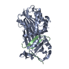


 PDBj
PDBj
 Assembly

 Sample preparation
 / Beamline: PX7.2 / Wavelength: 1.448
  Processing