 Movie
Movie Controller

[English] 日本語
 Yorodumi
- PDB-4cyg: The structure of vanin-1: defining the link between metabolic dis... -
+ Open data

- Basic information
| Entry | Database: PDB / ID: 4cyg | |||||||||
|---|---|---|---|---|---|---|---|---|---|---|
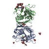
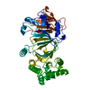
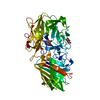
| Title | The structure of vanin-1: defining the link between metabolic disease, oxidative stress and inflammation | |||||||||
 Components Components | PANTETHEINASE | |||||||||
 Keywords Keywords |  HYDROLASE / INFLAMMATION / COLITIS / COA BIOSYNTHESIS / OXIDATIVE STRESS HYDROLASE / INFLAMMATION / COLITIS / COA BIOSYNTHESIS / OXIDATIVE STRESS | |||||||||
| Function / homology |  Function and homology informationpantetheine hydrolase / pantetheine hydrolase activity / pantothenate metabolic process / chronic inflammatory response / coenzyme A catabolic process / Vitamin B5 (pantothenate) metabolism / Post-translational modification: synthesis of GPI-anchored proteins / positive regulation of T cell differentiation in thymus / acute inflammatory response / positive regulation of oxidative stress-induced intrinsic apoptotic signaling pathway ...pantetheine hydrolase / pantetheine hydrolase activity / pantothenate metabolic process / chronic inflammatory response / coenzyme A catabolic process / Vitamin B5 (pantothenate) metabolism / Post-translational modification: synthesis of GPI-anchored proteins / positive regulation of T cell differentiation in thymus / acute inflammatory response / positive regulation of oxidative stress-induced intrinsic apoptotic signaling pathway / azurophil granule membrane / side of membrane / cell-cell adhesion / response to oxidative stress / inflammatory response / innate immune response / Neutrophil degranulation / extracellular region / membrane / plasma membrane Function and homology informationpantetheine hydrolase / pantetheine hydrolase activity / pantothenate metabolic process / chronic inflammatory response / coenzyme A catabolic process / Vitamin B5 (pantothenate) metabolism / Post-translational modification: synthesis of GPI-anchored proteins / positive regulation of T cell differentiation in thymus / acute inflammatory response / positive regulation of oxidative stress-induced intrinsic apoptotic signaling pathway ...pantetheine hydrolase / pantetheine hydrolase activity / pantothenate metabolic process / chronic inflammatory response / coenzyme A catabolic process / Vitamin B5 (pantothenate) metabolism / Post-translational modification: synthesis of GPI-anchored proteins / positive regulation of T cell differentiation in thymus / acute inflammatory response / positive regulation of oxidative stress-induced intrinsic apoptotic signaling pathway / azurophil granule membrane / side of membrane / cell-cell adhesion / response to oxidative stress / inflammatory response / innate immune response / Neutrophil degranulation / extracellular region / membrane / plasma membraneSimilarity search - Function | |||||||||
| Biological species |  HOMO SAPIENS (human) HOMO SAPIENS (human) | |||||||||
| Method | X-RAY DIFFRACTION / SYNCHROTRON / MOLECULAR REPLACEMENT / Resolution: 2.3 Å | |||||||||
 Authors Authors | Boersma, Y.L. / Newman, J. / Adams, T.E. / Sparrow, L. / Cowieson, N. / Lucent, D. / Krippner, G. / Bozaoglu, K. / Peat, T.S. | |||||||||
 Citation Citation | Journal: Acta Crystallogr.,Sect.D / Year: 2014 Title: The Structure of Vanin-1: A Key Enzyme Linking Metabolic Disease and Inflammation Authors: Boersma, Y.L. / Newman, J. / Adams, T.E. / Cowieson, N. / Krippner, G. / Bozaoglu, K. / Peat, T.S. | |||||||||
| History |
|
- Structure visualization
| Structure viewer | Molecule: MolmilJmol/JSmol |
|---|
- Downloads & links
-Download
| PDBx/mmCIF format | 4cyg.cif.gz | 205.2 KB | Display | PDBx/mmCIF format |
|---|---|---|---|---|
| PDB format | pdb4cyg.ent.gz | 163.1 KB | Display | PDB format |
| PDBx/mmJSON format | 4cyg.json.gz | Tree view | PDBx/mmJSON format | |
| Others |  Other downloads Other downloads |
-Validation report
| Arichive directory | https://data.pdbj.org/pub/pdb/validation_reports/cy/4cygftp://data.pdbj.org/pub/pdb/validation_reports/cy/4cyg | HTTPS FTP |
|---|
-Related structure data
| Related structure data | 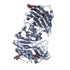 4cyfSC  4cyyC S: Starting model for refinement C: citing same article ( |
|---|---|
| Similar structure data |


-Links
 PDBj
PDBj- Assembly
| Deposited unit | 
| ||||||||
|---|---|---|---|---|---|---|---|---|---|
| 1 | 
| ||||||||
| 2 | 
| ||||||||
| Unit cell |
|
-Components
-Protein , 1 types, 2 molecules AB
| #1: Protein | Mass: 56325.219 Da / Num. of mol.: 2 Source method: isolated from a genetically manipulated source Details: FLAG TAG AT THE N-TERMINUS OF THE PROTEIN OTHERWISE THE PROTEIN IS THE MATURE FORM FOUND NATURALLY. Source: (gene. exp.) HOMO SAPIENS (human) / Description: DNA SYNTHESIZED / Cell line (production host): HUMAN EMBRYONIC KIDNEY / Production host: HOMO SAPIENS (human) / References: UniProt: O95497, pantetheine hydrolase |
|---|
-Sugars , 2 types, 7 molecules 
| #2: Polysaccharide | 2-acetamido-2-deoxy-beta-D-glucopyranose-(1-4)-2-acetamido-2-deoxy-beta-D-glucopyranose / Mass: 424.401 Da / Num. of mol.: 1 Source method: isolated from a genetically manipulated source |
|---|---|
| #4: Sugar | ChemComp-NAG / N-Acetylglucosamine Type: D-saccharide, beta linking / Mass: 221.208 Da / Num. of mol.: 6 Type: D-saccharide, beta linking / Mass: 221.208 Da / Num. of mol.: 6Source method: isolated from a genetically manipulated source Formula: C8H15NO6 |
-Non-polymers , 3 types, 311 molecules 


| #3: Chemical |  Mass: 295.374 Da / Num. of mol.: 2 / Source method: obtained synthetically / Formula: C16H25NO4 Mass: 295.374 Da / Num. of mol.: 2 / Source method: obtained synthetically / Formula: C16H25NO4#5: Chemical | Diethylene glycol Mass: 106.120 Da / Num. of mol.: 2 / Source method: obtained synthetically / Formula: C4H10O3 Mass: 106.120 Da / Num. of mol.: 2 / Source method: obtained synthetically / Formula: C4H10O3#6: Water | ChemComp-HOH / | WaterMass: 18.015 Da / Num. of mol.: 307 / Source method: isolated from a natural source / Formula: H2O |
|---|
-Details
| Sequence details | FLAG TAG AT N-TERMINUS INSTEAD OF SIGNAL PEPTIDE AND THE PRO-PEPTIDE AT THE C-TERMINUS HAS BEEN ...FLAG TAG AT N-TERMINUS INSTEAD OF SIGNAL PEPTIDE AND THE PRO-PEPTIDE AT THE C-TERMINUS HAS BEEN REMOVED TO GIVE THE MATURE PROTEIN. |
|---|
-Experimental details
-Experiment
| Experiment | Method: X-RAY DIFFRACTION / Number of used crystals: 1 |
|---|
- Sample preparation
| Crystal | Density Matthews: 3.88 Å3/Da / Density % sol: 68.3 % / Description: NONE |
|---|---|
| Crystal grow | Temperature: 281 K / Method: vapor diffusion, sitting drop / pH: 6.5 Details: CRYSTALLIZATION CONDITIONS: PROTEIN AT 12 MG/ML IN 50 MM BIS-TRIS PH 6.5 WITH 50 MM NACL. RESERVOIR CONDITIONS WERE 21% PEG 1500 WITH 10% (V/V) MALATE-MES-TRIS BUFFER AT PH 6.3. DROPS WERE ...Details: CRYSTALLIZATION CONDITIONS: PROTEIN AT 12 MG/ML IN 50 MM BIS-TRIS PH 6.5 WITH 50 MM NACL. RESERVOIR CONDITIONS WERE 21% PEG 1500 WITH 10% (V/V) MALATE-MES-TRIS BUFFER AT PH 6.3. DROPS WERE 200 NL PLUS 200 NL IN SITTING DROP PLATES AT 8C. |
-Data collection
| Diffraction | Mean temperature: 100 K |
|---|---|
| Diffraction source | Source: SYNCHROTRON / Site: Australian Synchrotron  / Beamline: MX2 / Wavelength: 0.9537 / Beamline: MX2 / Wavelength: 0.9537 |
| Detector | Type: ADSC CCD / Detector: CCD / Date: Nov 29, 2013 |
| Radiation | Protocol: SINGLE WAVELENGTH / Monochromatic (M) / Laue (L): M / Scattering type: x-ray |
| Radiation wavelength | Wavelength: 0.9537 Å / Relative weight: 1 |
| Reflection | Resolution: 2.3→48.3 Å / Num. obs: 72390 / % possible obs: 99.6 % / Observed criterion σ(I): 0 / Redundancy: 14.9 % / Rmerge(I) obs: 0.15 / Net I/σ(I): 15.6 |
| Reflection shell | Resolution: 2.3→2.42 Å / Redundancy: 15.2 % / Rmerge(I) obs: 1.24 / Mean I/σ(I) obs: 2.6 / % possible all: 99.3 |
- Processing
| Software |
| ||||||||||||||||||||||||||||||||||||||||||||||||||||||||||||||||||||||||||||||||||||||||||||||||||||||||||||||||||||||||||||||||||||||||||||||||||||||||||||||||||||||||||||||||||||||
|---|---|---|---|---|---|---|---|---|---|---|---|---|---|---|---|---|---|---|---|---|---|---|---|---|---|---|---|---|---|---|---|---|---|---|---|---|---|---|---|---|---|---|---|---|---|---|---|---|---|---|---|---|---|---|---|---|---|---|---|---|---|---|---|---|---|---|---|---|---|---|---|---|---|---|---|---|---|---|---|---|---|---|---|---|---|---|---|---|---|---|---|---|---|---|---|---|---|---|---|---|---|---|---|---|---|---|---|---|---|---|---|---|---|---|---|---|---|---|---|---|---|---|---|---|---|---|---|---|---|---|---|---|---|---|---|---|---|---|---|---|---|---|---|---|---|---|---|---|---|---|---|---|---|---|---|---|---|---|---|---|---|---|---|---|---|---|---|---|---|---|---|---|---|---|---|---|---|---|---|---|---|---|---|
| Refinement | Method to determine structure: MOLECULAR REPLACEMENT Starting model: PDB ENTRY 4CYF Resolution: 2.3→105.62 Å / Cor.coef. Fo:Fc: 0.953 / Cor.coef. Fo:Fc free: 0.935 / SU B: 5.191 / SU ML: 0.122 / Cross valid method: THROUGHOUT / ESU R: 0.2 / ESU R Free: 0.17 / Stereochemistry target values: MAXIMUM LIKELIHOOD Details: HYDROGENS HAVE BEEN ADDED IN THE RIDING POSITIONS. U VALUES REFINED INDIVIDUALLY THE RR6 INHIBITOR LOOKS TO BE COVALENTLY BOUND TO THE ACTIVE SITE CYS211 AS THE DENSITY IS CONTINUOUS IN THAT REGION.
| ||||||||||||||||||||||||||||||||||||||||||||||||||||||||||||||||||||||||||||||||||||||||||||||||||||||||||||||||||||||||||||||||||||||||||||||||||||||||||||||||||||||||||||||||||||||
| Solvent computation | Ion probe radii: 0.8 Å / Shrinkage radii: 0.8 Å / VDW probe radii: 1.2 Å / Solvent model: MASK | ||||||||||||||||||||||||||||||||||||||||||||||||||||||||||||||||||||||||||||||||||||||||||||||||||||||||||||||||||||||||||||||||||||||||||||||||||||||||||||||||||||||||||||||||||||||
| Displacement parameters | Biso mean: 36.863 Å2
| ||||||||||||||||||||||||||||||||||||||||||||||||||||||||||||||||||||||||||||||||||||||||||||||||||||||||||||||||||||||||||||||||||||||||||||||||||||||||||||||||||||||||||||||||||||||
| Refinement step | Cycle: LAST / Resolution: 2.3→105.62 Å
| ||||||||||||||||||||||||||||||||||||||||||||||||||||||||||||||||||||||||||||||||||||||||||||||||||||||||||||||||||||||||||||||||||||||||||||||||||||||||||||||||||||||||||||||||||||||
| Refine LS restraints |
|