 Movie
Movie Controller


+ Open data

- Basic information
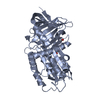
| Entry | Database: PDB / ID: 4caa | |||||||||
|---|---|---|---|---|---|---|---|---|---|---|
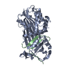
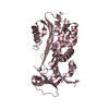
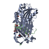
| Title | CLEAVED ANTICHYMOTRYPSIN T345R | |||||||||
 Components Components | (ANTICHYMOTRYPSIN) x 2 | |||||||||
 Keywords Keywords |  SERPIN / SERINE PROTEASE INHIBITOR / ANTICHYMOTRYPSIN SERPIN / SERINE PROTEASE INHIBITOR / ANTICHYMOTRYPSIN | |||||||||
| Function / homology |  Function and homology information Function and homology informationmaintenance of gastrointestinal epithelium / regulation of lipid metabolic process / platelet alpha granule lumen / acute-phase response / serine-type endopeptidase inhibitor activity / azurophil granule lumen / Platelet degranulation / secretory granule lumen / collagen-containing extracellular matrix / blood microparticle ...maintenance of gastrointestinal epithelium / regulation of lipid metabolic process / platelet alpha granule lumen / acute-phase response / serine-type endopeptidase inhibitor activity / azurophil granule lumen / Platelet degranulation / secretory granule lumen / collagen-containing extracellular matrix / blood microparticle / inflammatory response / Neutrophil degranulation / DNA binding / extracellular space / extracellular exosome / extracellular region / nucleusSimilarity search - Function | |||||||||
| Biological species |  Homo sapiens (human) Homo sapiens (human) | |||||||||
| Method | X-RAY DIFFRACTION / SYNCHROTRON / DIFFERENCE FOURIER / Resolution: 2.9 Å | |||||||||
 Authors Authors | Lukacs, C.M. / Christianson, D.W. | |||||||||
 Citation Citation | Journal: Biochemistry / Year: 1998 Title: Engineering an anion-binding cavity in antichymotrypsin modulates the "spring-loaded" serpin-protease interaction. Authors: Lukacs, C.M. / Rubin, H. / Christianson, D.W. #1: Journal: To be PublishedTitle: Burial of an Ion Pair in the Hydrophobic Core of Cleaved Ala-349->Arg Antichymotrypsin Compromises But Does not Obliterate Serpin Function Authors: Lukacs, C.M. / Rubin, H. / Christianson, D.W. | |||||||||
| History |
| |||||||||
| Remark 700 | SHEET RESIDUE A 345 IS MUTATION OF THR->ARG. THERE IS AN UNUSUAL TWIST TO THE BETA SHEET IN THIS ...SHEET RESIDUE A 345 IS MUTATION OF THR->ARG. THERE IS AN UNUSUAL TWIST TO THE BETA SHEET IN THIS AREA TO KEEP THE ARG SOLVENT EXPOSED. |
- Structure visualization
| Structure viewer | Molecule: MolmilJmol/JSmol |
|---|
- Downloads & links
-Download
| PDBx/mmCIF format | 4caa.cif.gz | 84.5 KB | Display | PDBx/mmCIF format |
|---|---|---|---|---|
| PDB format | pdb4caa.ent.gz | 63.9 KB | Display | PDB format |
| PDBx/mmJSON format | 4caa.json.gz | Tree view | PDBx/mmJSON format | |
| Others |  Other downloads Other downloads |
-Validation report
| Arichive directory | https://data.pdbj.org/pub/pdb/validation_reports/ca/4caaftp://data.pdbj.org/pub/pdb/validation_reports/ca/4caa | HTTPS FTP |
|---|
-Related structure data
| Related structure data |  1as4C 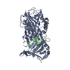 3caaC  1ct3 S: Starting model for refinement C: citing same article ( |
|---|---|
| Similar structure data |


-Links
 PDBj
PDBj
- Assembly
| Deposited unit | 
| ||||||||
|---|---|---|---|---|---|---|---|---|---|
| 1 |
| ||||||||
| Unit cell |
|
-Components
| #1: Protein | Mass: 38582.125 Da / Num. of mol.: 1 / Mutation: T345R Source method: isolated from a genetically manipulated source Details: CLEAVED ANTICHYMOTRYPSIN / Source: (gene. exp.) Homo sapiens (human) / Gene: ACT / Plasmid: PZMS / Gene (production host): ACT / Production host:  Escherichia coli (E. coli) / References: UniProt: P01011 Escherichia coli (E. coli) / References: UniProt: P01011 |
|---|---|
| #2: Protein/peptide | Mass: 4359.141 Da / Num. of mol.: 1 / Mutation: T345R Source method: isolated from a genetically manipulated source Details: CLEAVED ANTICHYMOTRYPSIN / Source: (gene. exp.) Homo sapiens (human) / Gene: ACT / Plasmid: PZMS / Gene (production host): ACT / Production host: Escherichia coli (E. coli) / References: UniProt: P01011 |
| Compound details | A: N-TERMINUS TO CLEAVAGE SITE (RESIDUES 20 - 358) B: CLEAVAGE SITE TO C-TERMINUS (RESIDUES 359 - 393) |
-Experimental details
-Experiment
| Experiment | Method: X-RAY DIFFRACTION / Number of used crystals: 1 |
|---|
- Sample preparation
| Crystal | Density Matthews: 3 Å3/Da / Density % sol: 59 % | ||||||||||||||||||||||||||||||||||||||||
|---|---|---|---|---|---|---|---|---|---|---|---|---|---|---|---|---|---|---|---|---|---|---|---|---|---|---|---|---|---|---|---|---|---|---|---|---|---|---|---|---|---|
| Crystal grow | pH: 5.6 Details: 14% PEG 8000 0.2 M MAGNESIUM ACETATE 0.1 M SODIUM CITRATE PH 5.6 | ||||||||||||||||||||||||||||||||||||||||
| Crystal | *PLUS | ||||||||||||||||||||||||||||||||||||||||
| Crystal grow | *PLUS Method: vapor diffusion, hanging drop / Details: Lukacs, C.M., (1996) Nat.Struct.Biol., 3, 888. | ||||||||||||||||||||||||||||||||||||||||
| Components of the solutions | *PLUS
|
-Data collection
| Diffraction | Mean temperature: 100 K |
|---|---|
| Diffraction source | Source: SYNCHROTRON / Site: CHESS  / Beamline: A1 / Wavelength: 1.54 / Beamline: A1 / Wavelength: 1.54 |
| Detector | Type: ADSC / Detector: CCD / Date: Sep 1, 1996 |
| Radiation | Monochromator: SI(111) / Monochromatic (M) / Laue (L): M / Scattering type: x-ray |
| Radiation wavelength | Wavelength: 1.54 Å / Relative weight: 1 |
| Reflection | Resolution: 2.9→20 Å / Num. obs: 37763 / % possible obs: 95.9 % / Redundancy: 3.35 % / Rsym value: 0.085 / Net I/σ(I): 7.1 |
| Reflection shell | Resolution: 2.9→3 Å / Redundancy: 3.3 % / Rsym value: 0.426 / % possible all: 97.1 |
| Reflection | *PLUS Num. obs: 11269 / Num. measured all: 37763 / Rmerge(I) obs: 0.085 |
- Processing
| Software |
| ||||||||||||||||||||||||||||||||||||||||||||||||||||||||||||
|---|---|---|---|---|---|---|---|---|---|---|---|---|---|---|---|---|---|---|---|---|---|---|---|---|---|---|---|---|---|---|---|---|---|---|---|---|---|---|---|---|---|---|---|---|---|---|---|---|---|---|---|---|---|---|---|---|---|---|---|---|---|
| Refinement | Method to determine structure: DIFFERENCE FOURIER Starting model: PDB ENTRY 1CT3 1ct3 Resolution: 2.9→8 Å / Rfactor Rfree error: 0.013 / Data cutoff high absF: 100000 / Data cutoff low absF: 0.1 / Cross valid method: THROUGHOUT / σ(F): 2 Details: GLN A 105 - ASP A 108 ARE IN VERY POOR ELECTRON DENSITY AND SHOULD BE TREATED AS SUCH. THEY HAVE BEEN REFINED WITH OCCUPANCIES OF 0.0. DATA WAS INDEXED WITH B>C IN ORDER TO USE DIFFERENCE ...Details: GLN A 105 - ASP A 108 ARE IN VERY POOR ELECTRON DENSITY AND SHOULD BE TREATED AS SUCH. THEY HAVE BEEN REFINED WITH OCCUPANCIES OF 0.0. DATA WAS INDEXED WITH B>C IN ORDER TO USE DIFFERENCE FOURIER TECHNIQUES FROM ENTRY 1CT3. THE DIFFERENCE IN UNIT CELLS IS PRESUMABLY DUE TO CRYOGENIC TECHNIQUE USED IN THIS DATA SET.
| ||||||||||||||||||||||||||||||||||||||||||||||||||||||||||||
| Displacement parameters | Biso mean: 35.5 Å2 | ||||||||||||||||||||||||||||||||||||||||||||||||||||||||||||
| Refinement step | Cycle: LAST / Resolution: 2.9→8 Å
| ||||||||||||||||||||||||||||||||||||||||||||||||||||||||||||
| Refine LS restraints |
| ||||||||||||||||||||||||||||||||||||||||||||||||||||||||||||
| LS refinement shell | Resolution: 2.9→3.03 Å / Rfactor Rfree error: 0.067 / Total num. of bins used: 8
| ||||||||||||||||||||||||||||||||||||||||||||||||||||||||||||
| Xplor file |
| ||||||||||||||||||||||||||||||||||||||||||||||||||||||||||||
| Software | *PLUS Name: X-PLOR / Version: 3.1 / Classification: refinement | ||||||||||||||||||||||||||||||||||||||||||||||||||||||||||||
| Refine LS restraints | *PLUS
|
