 Movie
Movie Controller

[English] 日本語
 Yorodumi
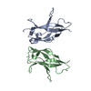
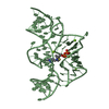
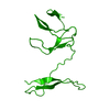
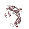
- PDB-1q8m: Crystal structure of the human myeloid cell activating receptor TREM-1 -
+ Open data

- Basic information
| Entry | Database: PDB / ID: 1q8m | ||||||
|---|---|---|---|---|---|---|---|
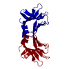
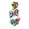
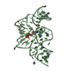
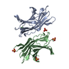
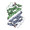
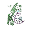
| Title | Crystal structure of the human myeloid cell activating receptor TREM-1 | ||||||
 Components Components | triggering receptor expressed on myeloid cells 1 TREM1 TREM1 | ||||||
 Keywords Keywords | IMMUNE SYSTEM RECEPTOR / V-type Ig-like domain / Immunoglobulin-like | ||||||
| Function / homology |  Function and homology information Function and homology informationreceptor decoy activity / neutrophil-mediated killing of gram-negative bacterium / humoral immune response / neutrophil chemotaxis / DAP12 interactions / Cell surface interactions at the vascular wall / Immunoregulatory interactions between a Lymphoid and a non-Lymphoid cell / transmembrane signaling receptor activity / signaling receptor activity / scaffold protein binding ...receptor decoy activity / neutrophil-mediated killing of gram-negative bacterium / humoral immune response / neutrophil chemotaxis / DAP12 interactions / Cell surface interactions at the vascular wall / Immunoregulatory interactions between a Lymphoid and a non-Lymphoid cell / transmembrane signaling receptor activity / signaling receptor activity / scaffold protein binding / adaptive immune response / intracellular signal transduction / innate immune response / cell surface / extracellular space / extracellular region / plasma membraneSimilarity search - Function | ||||||
| Biological species |  Homo sapiens (human) Homo sapiens (human) | ||||||
| Method | X-RAY DIFFRACTION / SYNCHROTRON / MAD / Resolution: 2.6 Å | ||||||
 Authors Authors | Radaev, S. / Kattah, M. / Rostro, B. / Colonna, M. / Sun, P.D. | ||||||
 Citation Citation | Journal: Structure / Year: 2003 Title: Crystal structure of the human myeloid cell activating receptor TREM-1 Authors: Radaev, S. / Kattah, M. / Rostro, B. / Colonna, M. / Sun, P.D. | ||||||
| History |
|
- Structure visualization
| Structure viewer | Molecule: MolmilJmol/JSmol |
|---|
- Downloads & links
-Download
| PDBx/mmCIF format | 1q8m.cif.gz | 110 KB | Display | PDBx/mmCIF format |
|---|---|---|---|---|
| PDB format | pdb1q8m.ent.gz | 89.2 KB | Display | PDB format |
| PDBx/mmJSON format | 1q8m.json.gz | Tree view | PDBx/mmJSON format | |
| Others |  Other downloads Other downloads |
-Validation report
| Arichive directory | https://data.pdbj.org/pub/pdb/validation_reports/q8/1q8mftp://data.pdbj.org/pub/pdb/validation_reports/q8/1q8m | HTTPS FTP |
|---|
-Related structure data
| Related structure data | |
|---|---|
| Similar structure data |


-Links
 PDBj
PDBj

- Assembly
| Deposited unit | 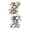
| ||||||||
|---|---|---|---|---|---|---|---|---|---|
| 1 | 
| ||||||||
| 2 | 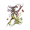
| ||||||||
| Unit cell |
|
-Components
| #1: Protein | TREM1 Mass: 14952.163 Da / Num. of mol.: 4 / Fragment: Extracellular domain Source method: isolated from a genetically manipulated source Source: (gene. exp.) Homo sapiens (human) / Gene: TREM1 / Plasmid: Pet-22b / Production host:  Escherichia coli (E. coli) / References: UniProt: Q9NP99 Escherichia coli (E. coli) / References: UniProt: Q9NP99#2: Chemical | ChemComp-SO4 / Sulfate  Mass: 96.063 Da / Num. of mol.: 10 / Source method: obtained synthetically / Formula: SO4 Mass: 96.063 Da / Num. of mol.: 10 / Source method: obtained synthetically / Formula: SO4#3: Chemical | ChemComp-GSH / | Glutathione  Mass: 307.323 Da / Num. of mol.: 1 / Source method: obtained synthetically / Formula: C10H17N3O6S Mass: 307.323 Da / Num. of mol.: 1 / Source method: obtained synthetically / Formula: C10H17N3O6S#4: Water | ChemComp-HOH / | Water Mass: 18.015 Da / Num. of mol.: 159 / Source method: isolated from a natural source / Formula: H2O Mass: 18.015 Da / Num. of mol.: 159 / Source method: isolated from a natural source / Formula: H2O |
|---|
-Experimental details
-Experiment
| Experiment | Method: X-RAY DIFFRACTION / Number of used crystals: 1 |
|---|
- Sample preparation
| Crystal | Density Matthews: 3.3 Å3/Da / Density % sol: 62.76 % | ||||||||||||||||||||||||
|---|---|---|---|---|---|---|---|---|---|---|---|---|---|---|---|---|---|---|---|---|---|---|---|---|---|
| Crystal grow | Temperature: 298 K / Method: vapor diffusion, hanging drop / pH: 5.7 Details: Ammonium sulfate, MES, pH 5.7, VAPOR DIFFUSION, HANGING DROP, temperature 298.0K | ||||||||||||||||||||||||
| Crystal grow | *PLUS Method: vapor diffusion, hanging drop / PH range low: 5.7 / PH range high: 5.4 | ||||||||||||||||||||||||
| Components of the solutions | *PLUS
|
-Data collection
| Diffraction | Mean temperature: 100 K | |||||||||||||||
|---|---|---|---|---|---|---|---|---|---|---|---|---|---|---|---|---|
| Diffraction source | Source: SYNCHROTRON / Site: APS  / Beamline: 19-ID / Wavelength: 1.0376, 0.9641, 0.9790, 0.9791 / Beamline: 19-ID / Wavelength: 1.0376, 0.9641, 0.9790, 0.9791 | |||||||||||||||
| Detector | Detector: CCD | |||||||||||||||
| Radiation | Protocol: SINGLE WAVELENGTH / Monochromatic (M) / Laue (L): M / Scattering type: x-ray | |||||||||||||||
| Radiation wavelength |
| |||||||||||||||
| Reflection | Resolution: 2.6→46 Å / Num. all: 52139 / Num. obs: 52139 / Observed criterion σ(I): -3 / Redundancy: 4.2 % / Biso Wilson estimate: 47 Å2 / Rsym value: 0.086 / Net I/σ(I): 23.5 | |||||||||||||||
| Reflection shell | Resolution: 2.6→2.7 Å / Redundancy: 4.2 % / Mean I/σ(I) obs: 4.2 / Num. unique all: 2369 / Rsym value: 0.44 / % possible all: 95.9 | |||||||||||||||
| Reflection | *PLUS Num. obs: 23897 / % possible obs: 98.6 % / Rmerge(I) obs: 0.086 | |||||||||||||||
| Reflection shell | *PLUS % possible obs: 97.7 % / Num. unique obs: 2369 / Rmerge(I) obs: 0.44 |
- Processing
| Software |
| ||||||||||||||||||||
|---|---|---|---|---|---|---|---|---|---|---|---|---|---|---|---|---|---|---|---|---|---|
| Refinement | Method to determine structure: MAD / Resolution: 2.6→46 Å / σ(F): 0 / Stereochemistry target values: Engh & Huber
| ||||||||||||||||||||
| Displacement parameters | Biso mean: 38 Å2 | ||||||||||||||||||||
| Refinement step | Cycle: LAST / Resolution: 2.6→46 Å
| ||||||||||||||||||||
| Refine LS restraints |
| ||||||||||||||||||||
| LS refinement shell | Resolution: 2.6→2.64 Å /
| ||||||||||||||||||||
| Refinement | *PLUS Lowest resolution: 46 Å / % reflection Rfree: 5 % | ||||||||||||||||||||
| Solvent computation | *PLUS | ||||||||||||||||||||
| Displacement parameters | *PLUS |
