 Movie
Movie Controller


+ Open data

- Basic information
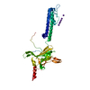
| Entry | Database: PDB / ID: 1i85 | ||||||
|---|---|---|---|---|---|---|---|
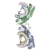
| Title | CRYSTAL STRUCTURE OF THE CTLA-4/B7-2 COMPLEX | ||||||
 Components Components |
| ||||||
 Keywords Keywords |  IMMUNE SYSTEM / Ig V-type domain IMMUNE SYSTEM / Ig V-type domain | ||||||
| Function / homology |  Function and homology information Function and homology informationpositive regulation of lymphotoxin A production / protein complex involved in cell adhesion / CD40 signaling pathway / negative regulation of regulatory T cell differentiation / positive regulation of T-helper 2 cell differentiation / activation of protein kinase C activity / RUNX1 and FOXP3 control the development of regulatory T lymphocytes (Tregs) / clathrin-coated endocytic vesicle / CD28 co-stimulation / CD28 dependent Vav1 pathway ...positive regulation of lymphotoxin A production / protein complex involved in cell adhesion / CD40 signaling pathway / negative regulation of regulatory T cell differentiation / positive regulation of T-helper 2 cell differentiation / activation of protein kinase C activity / RUNX1 and FOXP3 control the development of regulatory T lymphocytes (Tregs) / clathrin-coated endocytic vesicle / CD28 co-stimulation / CD28 dependent Vav1 pathway / positive regulation of immunoglobulin production / CTLA4 inhibitory signaling / B cell activation / positive regulation of interleukin-4 production / negative regulation of B cell proliferation / Interleukin-10 signaling / CD28 dependent PI3K/Akt signaling / : / centriolar satellite / coreceptor activity / negative regulation of T cell proliferation / positive regulation of T cell proliferation / T cell costimulation / T cell activation / positive regulation of interleukin-2 production / B cell receptor signaling pathway / positive regulation of non-canonical NF-kappaB signal transduction / Constitutive Signaling by Aberrant PI3K in Cancer / virus receptor activity / PIP3 activates AKT signaling / signaling receptor activity / T cell receptor signaling pathway / PI5P, PP2A and IER3 Regulate PI3K/AKT Signaling / adaptive immune response / cellular response to lipopolysaccharide / receptor ligand activity / cell surface receptor signaling pathway / immune response / positive regulation of apoptotic process / external side of plasma membrane / DNA damage response / positive regulation of cell population proliferation / perinuclear region of cytoplasm / positive regulation of DNA-templated transcription / Golgi apparatus / cell surface / extracellular exosome / plasma membraneSimilarity search - Function | ||||||
| Biological species |  Homo sapiens (human) Homo sapiens (human) | ||||||
| Method | X-RAY DIFFRACTION / SYNCHROTRON / MOLECULAR REPLACEMENT / Resolution: 3.2 Å | ||||||
 Authors Authors | Schwartz, J.-C.D. / Zhang, X. / Fedorov, A.A. / Nathenson, S.G. / Almo, S.C. | ||||||
 Citation Citation | Journal: Nature / Year: 2001 Title: Structural basis for co-stimulation by the human CTLA-4/B7-2 complex. Authors: Schwartz, J.C. / Zhang, X. / Fedorov, A.A. / Nathenson, S.G. / Almo, S.C. | ||||||
| History |
|
- Structure visualization
| Structure viewer | Molecule: MolmilJmol/JSmol |
|---|
- Downloads & links
-Download
| PDBx/mmCIF format | 1i85.cif.gz | 92.8 KB | Display | PDBx/mmCIF format |
|---|---|---|---|---|
| PDB format | pdb1i85.ent.gz | 77.2 KB | Display | PDB format |
| PDBx/mmJSON format | 1i85.json.gz | Tree view | PDBx/mmJSON format | |
| Others |  Other downloads Other downloads |
-Validation report
| Arichive directory | https://data.pdbj.org/pub/pdb/validation_reports/i8/1i85ftp://data.pdbj.org/pub/pdb/validation_reports/i8/1i85 | HTTPS FTP |
|---|
-Related structure data
| Similar structure data |
|---|


-Links
 PDBj
PDBj


- Assembly
| Deposited unit | 
| ||||||||
|---|---|---|---|---|---|---|---|---|---|
| 1 |
| ||||||||
| Unit cell |
| ||||||||
| Details | The CTLA-4 homodimer is generated by Chain D and the translatonal symmetry mate of chain C (apply the operator x,y,z+1 to Chain C) / The repeating arrangement of CTLA-4 and B7-2 dimers is generated by applying the translational symmetry operation (x,y,z+/-N; where N ranges over all integers) to all chains in the asymmetric unit |
-Components
| #1: Antibody | Mass: 12849.631 Da / Num. of mol.: 2 / Fragment: IG V-TYPE (RECEPTOR BINDING) DOMAIN Source method: isolated from a genetically manipulated source Source: (gene. exp.) Homo sapiens (human) / Gene: CD86 / Plasmid: PET3A / Species (production host): Escherichia coli / Production host:  Escherichia coli BL21(DE3) (bacteria) / Strain (production host): BL21 (DE3) / References: UniProt: P42081 Escherichia coli BL21(DE3) (bacteria) / Strain (production host): BL21 (DE3) / References: UniProt: P42081#2: Protein | Mass: 13510.340 Da / Num. of mol.: 2 / Fragment: EXTRACELLULAR DOMAIN Source method: isolated from a genetically manipulated source Source: (gene. exp.) Homo sapiens (human) / Gene: CTLA4 / Plasmid: PET3A / Species (production host): Escherichia coli / Production host: Escherichia coli BL21(DE3) (bacteria) / Strain (production host): BL21 (DE3) / References: UniProt: P16410 |
|---|
-Experimental details
-Experiment
| Experiment | Method: X-RAY DIFFRACTION / Number of used crystals: 1 |
|---|
- Sample preparation
| Crystal | Density Matthews: 2.59 Å3/Da / Density % sol: 52 % | ||||||||||||||||||||
|---|---|---|---|---|---|---|---|---|---|---|---|---|---|---|---|---|---|---|---|---|---|
| Crystal grow | Temperature: 298 K / Method: vapor diffusion, hanging drop / pH: 7 Details: PEG20K, HEPES, pH 7.0, VAPOR DIFFUSION, HANGING DROP, temperature 298K | ||||||||||||||||||||
| Crystal grow | *PLUS Details: drop consists of equal amounts of protein and reservoir solutions | ||||||||||||||||||||
| Components of the solutions | *PLUS
|
-Data collection
| Diffraction | Mean temperature: 100 K |
|---|---|
| Diffraction source | Source: SYNCHROTRON / Site: NSLS  / Beamline: X9B / Wavelength: 0.98 Å / Beamline: X9B / Wavelength: 0.98 Å |
| Detector | Type: ADSC QUANTUM 4 / Detector: CCD / Date: Jan 1, 2000 |
| Radiation | Monochromator: NULL / Protocol: SINGLE WAVELENGTH / Monochromatic (M) / Laue (L): M / Scattering type: x-ray |
| Radiation wavelength | Wavelength: 0.98 Å / Relative weight: 1 |
| Reflection | Resolution: 3.2→25 Å / Num. all: 7080 / Num. obs: 7080 / % possible obs: 78.8 % / Observed criterion σ(F): 0 / Observed criterion σ(I): 0 / Redundancy: 2.4 % / Rmerge(I) obs: 0.121 / Net I/σ(I): 6.6 |
| Reflection shell | Resolution: 3.2→3.31 Å / Redundancy: 2.3 % / Rmerge(I) obs: 0.321 / Mean I/σ(I) obs: 2.2 / % possible all: 79.4 |
| Reflection shell | *PLUS % possible obs: 79.4 % |
- Processing
| Software |
| |||||||||||||||||||||||||
|---|---|---|---|---|---|---|---|---|---|---|---|---|---|---|---|---|---|---|---|---|---|---|---|---|---|---|
| Refinement | Method to determine structure: MOLECULAR REPLACEMENT / Resolution: 3.2→10 Å / Cross valid method: THROUGHOUT / σ(F): 1.2 / σ(I): 1.4 / Stereochemistry target values: Engh & Huber
| |||||||||||||||||||||||||
| Displacement parameters | Biso mean: 20.4 Å2 | |||||||||||||||||||||||||
| Refine analyze |
| |||||||||||||||||||||||||
| Refinement step | Cycle: LAST / Resolution: 3.2→10 Å
| |||||||||||||||||||||||||
| Refine LS restraints |
| |||||||||||||||||||||||||
| LS refinement shell | Resolution: 3.2→3.31 Å / Total num. of bins used: 10
| |||||||||||||||||||||||||
| Xplor file | Serial no: 1 / Param file: protein_rep.param / Topol file: protein.top | |||||||||||||||||||||||||
| Software | *PLUS Name: CNS / Version: 0.9 / Classification: refinement | |||||||||||||||||||||||||
| Refine LS restraints | *PLUS
|
