 Movie
Movie Controller

[English] 日本語
 Yorodumi
- PDB-3ocp: Crystal structure of cAMP bound cGMP-dependent protein kinase(92-227) -
+ Open data

- Basic information
| Entry | Database: PDB / ID: 3ocp | ||||||
|---|---|---|---|---|---|---|---|
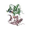
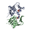
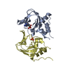
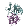
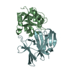
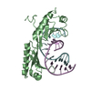
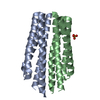
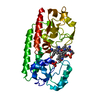
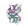
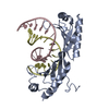
| Title | Crystal structure of cAMP bound cGMP-dependent protein kinase(92-227) | ||||||
 Components Components | PRKG1 protein | ||||||
 Keywords Keywords | TRANSFERASE / serine/threonine kinase / TF2I and IRAG | ||||||
| Function / homology |  Function and homology information Function and homology informationnegative regulation of inositol phosphate biosynthetic process / negative regulation of glutamate secretion / cGMP-dependent protein kinase / cGMP-dependent protein kinase activity / cell growth involved in cardiac muscle cell development / regulation of testosterone biosynthetic process / collateral sprouting / negative regulation of vascular associated smooth muscle cell migration / negative regulation of platelet aggregation / relaxation of vascular associated smooth muscle ...negative regulation of inositol phosphate biosynthetic process / negative regulation of glutamate secretion / cGMP-dependent protein kinase / cGMP-dependent protein kinase activity / cell growth involved in cardiac muscle cell development / regulation of testosterone biosynthetic process / collateral sprouting / negative regulation of vascular associated smooth muscle cell migration / negative regulation of platelet aggregation / relaxation of vascular associated smooth muscle / positive regulation of circadian rhythm / Rap1 signalling / mitogen-activated protein kinase p38 binding / regulation of GTPase activity / dendrite development / spermatid development / cGMP effects / negative regulation of vascular associated smooth muscle cell proliferation / cGMP-mediated signaling / calcium channel regulator activity / cGMP binding / forebrain development / cerebellum development / acrosomal vesicle / neuron migration / sarcolemma / Ca2+ pathway / positive regulation of cytosolic calcium ion concentration / actin cytoskeleton organization / protein kinase activity / protein phosphorylation / protein serine kinase activity / Golgi apparatus / signal transduction / nucleoplasm / ATP binding / identical protein binding / plasma membrane / cytosol / cytoplasmSimilarity search - Function | ||||||
| Biological species |  Homo sapiens (human) Homo sapiens (human) | ||||||
| Method | X-RAY DIFFRACTION / SYNCHROTRON / MOLECULAR REPLACEMENT / Resolution: 2.49 Å | ||||||
 Authors Authors | Kim, J.J. / Huang, G. / Kwon, T.K. / Zwart, P. / Headd, J. / Kim, C. | ||||||
 Citation Citation | Journal: Plos One / Year: 2011 Title: Co-Crystal Structures of PKG Ibeta (92-227) with cGMP and cAMP Reveal the Molecular Details of Cyclic-Nucleotide Binding Authors: Kim, J.J. / Casteel, D.E. / Huang, G. / Kwon, T.H. / Ren, R.K. / Zwart, P. / Headd, J.J. / Brown, N.G. / Chow, D.C. / Palzkill, T. / Kim, C. | ||||||
| History |
|
- Structure visualization
| Structure viewer | Molecule: MolmilJmol/JSmol |
|---|
- Downloads & links
-Download
| PDBx/mmCIF format | 3ocp.cif.gz | 66 KB | Display | PDBx/mmCIF format |
|---|---|---|---|---|
| PDB format | pdb3ocp.ent.gz | 48.1 KB | Display | PDB format |
| PDBx/mmJSON format | 3ocp.json.gz | Tree view | PDBx/mmJSON format | |
| Others |  Other downloads Other downloads |
-Validation report
| Arichive directory | https://data.pdbj.org/pub/pdb/validation_reports/oc/3ocpftp://data.pdbj.org/pub/pdb/validation_reports/oc/3ocp | HTTPS FTP |
|---|
-Related structure data
| Related structure data |  3od0C  3ogjC  1rgsS C: citing same article ( S: Starting model for refinement |
|---|---|
| Similar structure data |
-Links
 PDBj
PDBj


- Assembly
| Deposited unit | 
| ||||||||
|---|---|---|---|---|---|---|---|---|---|
| 1 |
| ||||||||
| Unit cell |
|
-Components
| #1: Protein | Mass: 15607.178 Da / Num. of mol.: 2 / Fragment: Cyclic nucleotie binding domain Source method: isolated from a genetically manipulated source Source: (gene. exp.) Homo sapiens (human) / Plasmid: pQTEV / Production host:  Escherichia coli (E. coli) / Strain (production host): BL21(DE3) Escherichia coli (E. coli) / Strain (production host): BL21(DE3)References: UniProt: Q6P5T7, UniProt: Q13976*PLUS, cGMP-dependent protein kinase#2: Chemical | Cyclic adenosine monophosphate  Mass: 329.206 Da / Num. of mol.: 2 / Source method: obtained synthetically / Formula: C10H12N5O6P Mass: 329.206 Da / Num. of mol.: 2 / Source method: obtained synthetically / Formula: C10H12N5O6P#3: Water | ChemComp-HOH / | Water Mass: 18.015 Da / Num. of mol.: 99 / Source method: isolated from a natural source / Formula: H2O Mass: 18.015 Da / Num. of mol.: 99 / Source method: isolated from a natural source / Formula: H2O |
|---|
-Experimental details
-Experiment
| Experiment | Method: X-RAY DIFFRACTION / Number of used crystals: 1 |
|---|
- Sample preparation
| Crystal | Density Matthews: 4.48 Å3/Da / Density % sol: 72.57 % |
|---|---|
| Crystal grow | Temperature: 277.15 K / Method: vapor diffusion, hanging drop / pH: 5.6 Details: 1.4 M sodium/potassium phosphate, pH 5.6, VAPOR DIFFUSION, HANGING DROP, temperature 277.15K |
-Data collection
| Diffraction | Mean temperature: 100 K |
|---|---|
| Diffraction source | Source: SYNCHROTRON / Site: ALS  / Beamline: 5.0.1 / Wavelength: 1 Å / Beamline: 5.0.1 / Wavelength: 1 Å |
| Detector | Type: ADSC QUANTUM 315r / Detector: CCD / Date: Dec 22, 2009 |
| Radiation | Monochromator: Si(220) Asymmetric cut single crystal / Protocol: SINGLE WAVELENGTH / Monochromatic (M) / Laue (L): M / Scattering type: x-ray |
| Radiation wavelength | Wavelength: 1 Å / Relative weight: 1 |
| Reflection | Resolution: 2.49→50 Å / Num. all: 20607 / Num. obs: 20339 / % possible obs: 98.7 % / Observed criterion σ(F): 1 / Observed criterion σ(I): 1 / Redundancy: 13.8 % / Rmerge(I) obs: 0.105 / Rsym value: 0.101 |
- Processing
| Software |
| ||||||||||||||||||||||||||||||||||||||||||||||||||||||||
|---|---|---|---|---|---|---|---|---|---|---|---|---|---|---|---|---|---|---|---|---|---|---|---|---|---|---|---|---|---|---|---|---|---|---|---|---|---|---|---|---|---|---|---|---|---|---|---|---|---|---|---|---|---|---|---|---|---|
| Refinement | Method to determine structure: MOLECULAR REPLACEMENT Starting model: PDB ENTRY 1RGS Resolution: 2.49→46.393 Å / SU ML: 0.27 / σ(F): 0 / σ(I): 2 / Stereochemistry target values: ML
| ||||||||||||||||||||||||||||||||||||||||||||||||||||||||
| Solvent computation | Shrinkage radii: 0.72 Å / VDW probe radii: 1 Å / Solvent model: FLAT BULK SOLVENT MODEL / Bsol: 60 Å2 / ksol: 0.374 e/Å3 | ||||||||||||||||||||||||||||||||||||||||||||||||||||||||
| Displacement parameters |
| ||||||||||||||||||||||||||||||||||||||||||||||||||||||||
| Refinement step | Cycle: LAST / Resolution: 2.49→46.393 Å
| ||||||||||||||||||||||||||||||||||||||||||||||||||||||||
| Refine LS restraints |
| ||||||||||||||||||||||||||||||||||||||||||||||||||||||||
| LS refinement shell |
|
