 Movie
Movie Controller


+ Open data

- Basic information
| Entry | Database: PDB / ID: 3jss | ||||||
|---|---|---|---|---|---|---|---|
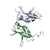
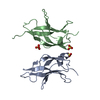
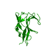
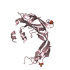
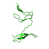
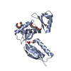
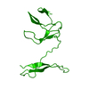
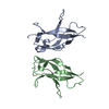
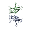
| Title | Crystal structure of a mutant RelB dimerization domain | ||||||
 Components Components | Transcription factor RelB | ||||||
 Keywords Keywords |  TRANSCRIPTION / NF-kB / intertwined dimer / Activator / Nucleus / Phosphoprotein / Transcription regulation TRANSCRIPTION / NF-kB / intertwined dimer / Activator / Nucleus / Phosphoprotein / Transcription regulation | ||||||
| Function / homology |  Function and homology information Function and homology informationT-helper 1 cell differentiation / Dectin-1 mediated noncanonical NF-kB signaling / NIK-->noncanonical NF-kB signaling / CD209 (DC-SIGN) signaling / myeloid dendritic cell differentiation / negative regulation of interferon-beta production / cellular response to osmotic stress / T-helper 1 type immune response / non-canonical NF-kappaB signal transduction / antigen processing and presentation ...T-helper 1 cell differentiation / Dectin-1 mediated noncanonical NF-kB signaling / NIK-->noncanonical NF-kB signaling / CD209 (DC-SIGN) signaling / myeloid dendritic cell differentiation / negative regulation of interferon-beta production / cellular response to osmotic stress / T-helper 1 type immune response / non-canonical NF-kappaB signal transduction / antigen processing and presentation / canonical NF-kappaB signal transduction / transcription repressor complex / response to cytokine / circadian regulation of gene expression / DNA-binding transcription factor activity, RNA polymerase II-specific / inflammatory response / RNA polymerase II cis-regulatory region sequence-specific DNA binding / DNA-binding transcription factor activity / innate immune response / negative regulation of DNA-templated transcription / centrosome / synapse / chromatin / protein kinase binding / positive regulation of transcription by RNA polymerase II / DNA binding / nucleoplasm / identical protein binding / nucleus / cytosolSimilarity search - Function | ||||||
| Biological species |  Mus musculus (house mouse) Mus musculus (house mouse) | ||||||
| Method | X-RAY DIFFRACTION / MOLECULAR REPLACEMENT / molecular replacement / Resolution: 2.6 Å | ||||||
 Authors Authors | Vu, D. / Huang, D.B. / Ghosh, G. | ||||||
 Citation Citation | Journal: J.Mol.Biol. / Year: 2013 Title: A structural basis for selective dimerization by NF-kappa B RelB. Authors: Vu, D. / Huang, D.B. / Vemu, A. / Ghosh, G. | ||||||
| History |
|
- Structure visualization
| Structure viewer | Molecule: MolmilJmol/JSmol |
|---|
- Downloads & links
-Download
| PDBx/mmCIF format | 3jss.cif.gz | 31.3 KB | Display | PDBx/mmCIF format |
|---|---|---|---|---|
| PDB format | pdb3jss.ent.gz | 20.5 KB | Display | PDB format |
| PDBx/mmJSON format | 3jss.json.gz | Tree view | PDBx/mmJSON format | |
| Others |  Other downloads Other downloads |
-Validation report
| Arichive directory | https://data.pdbj.org/pub/pdb/validation_reports/js/3jssftp://data.pdbj.org/pub/pdb/validation_reports/js/3jss | HTTPS FTP |
|---|
-Related structure data


-Links
 PDBj
PDBj


- Assembly
| Deposited unit | 
| ||||||||
|---|---|---|---|---|---|---|---|---|---|
| 1 | 
| ||||||||
| Unit cell |
|
-Components
| #1: Protein | Mass: 11460.842 Da / Num. of mol.: 1 / Fragment: dimerization domain (UNP residues 278-378) / Mutation: V314R, A324G, F358Q, L362K Source method: isolated from a genetically manipulated source Source: (gene. exp.) Mus musculus (house mouse) / Gene: RelB / Plasmid: PET15B / Production host:  Escherichia coli (E. coli) / Strain (production host): BL21 / References: UniProt: Q04863 Escherichia coli (E. coli) / Strain (production host): BL21 / References: UniProt: Q04863 |
|---|
-Experimental details
-Experiment
| Experiment | Method: X-RAY DIFFRACTION / Number of used crystals: 1 |
|---|
- Sample preparation
| Crystal | Density Matthews: 3.94 Å3/Da / Density % sol: 68.82 % |
|---|---|
| Crystal grow | Temperature: 291 K / Method: vapor diffusion, hanging drop / pH: 6.5 Details: 20% PEG800, 0.1M ammonium sulfate, pH 6.5, VAPOR DIFFUSION, HANGING DROP, temperature 291K |
-Data collection
| Diffraction | Mean temperature: 105 K |
|---|---|
| Diffraction source | Source: ROTATING ANODE / Type: RIGAKU RU200 / Wavelength: 1.5418 Å |
| Detector | Type: MAR scanner 345 mm plate / Detector: IMAGE PLATE / Date: Jan 1, 2006 / Details: mirros |
| Radiation | Protocol: SINGLE WAVELENGTH / Monochromatic (M) / Laue (L): M / Scattering type: x-ray |
| Radiation wavelength | Wavelength: 1.5418 Å / Relative weight: 1 |
| Reflection | Resolution: 2.6→30 Å / Num. all: 5318 / Num. obs: 4987 / % possible obs: 85 % / Observed criterion σ(I): 1 / Redundancy: 12 % / Rmerge(I) obs: 0.073 / Net I/σ(I): 14.7 |
| Reflection shell | Resolution: 2.6→2.69 Å / Redundancy: 10 % / Rmerge(I) obs: 0.29 / Mean I/σ(I) obs: 2.8 / Num. unique all: 288 / % possible all: 50 |
-Phasing
| Phasing | Method: molecular replacement |
|---|
- Processing
| Software |
| ||||||||||||||||||||||||||||||||||||
|---|---|---|---|---|---|---|---|---|---|---|---|---|---|---|---|---|---|---|---|---|---|---|---|---|---|---|---|---|---|---|---|---|---|---|---|---|---|
| Refinement | Method to determine structure: MOLECULAR REPLACEMENT / Resolution: 2.6→23.48 Å / Rfactor Rfree error: 0.013 / Occupancy max: 1 / Occupancy min: 1 / Data cutoff high absF: 362431 / Data cutoff low absF: 0 / Isotropic thermal model: RESTRAINED / Cross valid method: THROUGHOUT / σ(F): 2
| ||||||||||||||||||||||||||||||||||||
| Solvent computation | Solvent model: FLAT MODEL / Bsol: 27.682 Å2 / ksol: 0.358 e/Å3 | ||||||||||||||||||||||||||||||||||||
| Displacement parameters | Biso max: 114.1 Å2 / Biso mean: 39.386 Å2 / Biso min: 7.71 Å2
| ||||||||||||||||||||||||||||||||||||
| Refine analyze |
| ||||||||||||||||||||||||||||||||||||
| Refinement step | Cycle: LAST / Resolution: 2.6→23.48 Å
| ||||||||||||||||||||||||||||||||||||
| Refine LS restraints |
| ||||||||||||||||||||||||||||||||||||
| LS refinement shell | Resolution: 2.6→2.76 Å / Rfactor Rfree error: 0.051 / Total num. of bins used: 6
| ||||||||||||||||||||||||||||||||||||
| Xplor file | Serial no: 1 / Param file: protein_rep.param / Topol file: protein.top |
