 Movie
Movie Controller

[English] 日本語
 Yorodumi
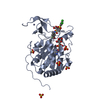
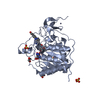
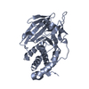
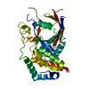
- PDB-1pa1: Crystal structure of the C215D mutant of protein tyrosine phospha... -
+ Open data

- Basic information
| Entry | Database: PDB / ID: 1pa1 | ||||||
|---|---|---|---|---|---|---|---|
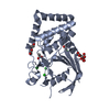
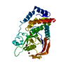
| Title | Crystal structure of the C215D mutant of protein tyrosine phosphatase 1B | ||||||
 Components Components | Protein-tyrosine phosphatase, non-receptor type 1 | ||||||
 Keywords Keywords |  HYDROLASE / phosphatase / catalytic loop / active-site mutant HYDROLASE / phosphatase / catalytic loop / active-site mutant | ||||||
| Function / homology |  Function and homology information Function and homology informationregulation of hepatocyte growth factor receptor signaling pathway / PTK6 Down-Regulation / peptidyl-tyrosine dephosphorylation involved in inactivation of protein kinase activity / positive regulation of receptor catabolic process / insulin receptor recycling / negative regulation of PERK-mediated unfolded protein response / negative regulation of vascular endothelial growth factor receptor signaling pathway / regulation of intracellular protein transport / IRE1-mediated unfolded protein response / cytoplasmic side of endoplasmic reticulum membrane ...regulation of hepatocyte growth factor receptor signaling pathway / PTK6 Down-Regulation / peptidyl-tyrosine dephosphorylation involved in inactivation of protein kinase activity / positive regulation of receptor catabolic process / insulin receptor recycling / negative regulation of PERK-mediated unfolded protein response / negative regulation of vascular endothelial growth factor receptor signaling pathway / regulation of intracellular protein transport / IRE1-mediated unfolded protein response / cytoplasmic side of endoplasmic reticulum membrane / platelet-derived growth factor receptor-beta signaling pathway / sorting endosome / mitochondrial crista / positive regulation of IRE1-mediated unfolded protein response / regulation of type I interferon-mediated signaling pathway / regulation of endocytosis / non-membrane spanning protein tyrosine phosphatase activity / positive regulation of protein tyrosine kinase activity / peptidyl-tyrosine dephosphorylation / Regulation of IFNA/IFNB signaling / regulation of signal transduction / cellular response to unfolded protein / growth hormone receptor signaling pathway via JAK-STAT / negative regulation of endoplasmic reticulum stress-induced intrinsic apoptotic signaling pathway / negative regulation of signal transduction / Regulation of IFNG signaling / Growth hormone receptor signaling / MECP2 regulates neuronal receptors and channels / endoplasmic reticulum unfolded protein response / positive regulation of JUN kinase activity / Insulin receptor recycling / negative regulation of insulin receptor signaling pathway / ephrin receptor binding / Integrin signaling / protein dephosphorylation / protein-tyrosine-phosphatase / protein phosphatase 2A binding / negative regulation of MAP kinase activity / protein tyrosine phosphatase activity / endosome lumen / insulin receptor binding / Negative regulation of MET activity / negative regulation of ERK1 and ERK2 cascade / receptor tyrosine kinase binding / insulin receptor signaling pathway / actin cytoskeleton organization / early endosome / mitochondrial matrix / cadherin binding / protein kinase binding / enzyme binding / endoplasmic reticulum / protein-containing complex / RNA binding / zinc ion binding / cytosol / cytoplasmSimilarity search - Function | ||||||
| Biological species |  Homo sapiens (human) Homo sapiens (human) | ||||||
| Method | X-RAY DIFFRACTION / SYNCHROTRON / FOURIER SYNTHESIS / Resolution: 1.6 Å | ||||||
 Authors Authors | Romsicki, Y. / Scapin, G. / Beaulieu-Audy, V. / Patel, S.B. / Becker, J.W. / Kennedy, B. / Asante-Appiah, E. | ||||||
 Citation Citation | Journal: J.Biol.Chem. / Year: 2003 Title: Functional characterization and crystal structure of the C215D mutant of protein-tyrosine phosphatase-1B Authors: Romsicki, Y. / Scapin, G. / Beaulieu-Audy, V. / Patel, S. / Becker, J.W. / Kennedy, B.P. / Asante-Appiah, E. | ||||||
| History |
|
- Structure visualization
| Structure viewer | Molecule: MolmilJmol/JSmol |
|---|
- Downloads & links
-Download
| PDBx/mmCIF format | 1pa1.cif.gz | 90.1 KB | Display | PDBx/mmCIF format |
|---|---|---|---|---|
| PDB format | pdb1pa1.ent.gz | 67.5 KB | Display | PDB format |
| PDBx/mmJSON format | 1pa1.json.gz | Tree view | PDBx/mmJSON format | |
| Others |  Other downloads Other downloads |
-Validation report
| Arichive directory | https://data.pdbj.org/pub/pdb/validation_reports/pa/1pa1ftp://data.pdbj.org/pub/pdb/validation_reports/pa/1pa1 | HTTPS FTP |
|---|
-Related structure data
| Related structure data |  1ptyS S: Starting model for refinement |
|---|---|
| Similar structure data |


-Links
 PDBj
PDBj


- Assembly
| Deposited unit | 
| ||||||||
|---|---|---|---|---|---|---|---|---|---|
| 1 |
| ||||||||
| Unit cell |
|
-Components
| #1: Protein | Mass: 36250.117 Da / Num. of mol.: 1 / Fragment: catalytic domain / Mutation: C215D Source method: isolated from a genetically manipulated source Source: (gene. exp.) Homo sapiens (human) / Species (production host): Escherichia coli / Production host:  Escherichia coli BL21(DE3) (bacteria) / Strain (production host): BL21(DE3) / References: UniProt: P18031, protein-tyrosine-phosphatase Escherichia coli BL21(DE3) (bacteria) / Strain (production host): BL21(DE3) / References: UniProt: P18031, protein-tyrosine-phosphatase | ||||
|---|---|---|---|---|---|
| #2: Chemical |   Mass: 24.305 Da / Num. of mol.: 2 / Source method: obtained synthetically / Formula: Mg Mass: 24.305 Da / Num. of mol.: 2 / Source method: obtained synthetically / Formula: Mg#3: Chemical | ChemComp-CL / Chloride  Mass: 35.453 Da / Num. of mol.: 6 / Source method: obtained synthetically / Formula: Cl Mass: 35.453 Da / Num. of mol.: 6 / Source method: obtained synthetically / Formula: Cl#4: Water | ChemComp-HOH / | Water Mass: 18.015 Da / Num. of mol.: 325 / Source method: isolated from a natural source / Formula: H2O Mass: 18.015 Da / Num. of mol.: 325 / Source method: isolated from a natural source / Formula: H2O |
-Experimental details
-Experiment
| Experiment | Method: X-RAY DIFFRACTION / Number of used crystals: 1 |
|---|
- Sample preparation
| Crystal | Density Matthews: 3.25 Å3/Da / Density % sol: 62.15 % | |||||||||||||||||||||||||||||||||||||||||||||||||||||||||||||||
|---|---|---|---|---|---|---|---|---|---|---|---|---|---|---|---|---|---|---|---|---|---|---|---|---|---|---|---|---|---|---|---|---|---|---|---|---|---|---|---|---|---|---|---|---|---|---|---|---|---|---|---|---|---|---|---|---|---|---|---|---|---|---|---|---|
| Crystal grow | Temperature: 277 K / Method: vapor diffusion, sitting drop / pH: 7 Details: PEG 3350, MgCl2, Hepes, pH 7.0, VAPOR DIFFUSION, SITTING DROP, temperature 277K | |||||||||||||||||||||||||||||||||||||||||||||||||||||||||||||||
| Crystal grow | *PLUS Temperature: 4 ℃ / Method: vapor diffusion, sitting drop | |||||||||||||||||||||||||||||||||||||||||||||||||||||||||||||||
| Components of the solutions | *PLUS
|
-Data collection
| Diffraction | Mean temperature: 100 K |
|---|---|
| Diffraction source | Source: SYNCHROTRON / Site: APS  / Beamline: 17-ID / Wavelength: 1 Å / Beamline: 17-ID / Wavelength: 1 Å |
| Detector | Type: ADSC QUANTUM 210 / Detector: CCD / Date: Jun 14, 2002 |
| Radiation | Monochromator: NA / Protocol: SINGLE WAVELENGTH / Monochromatic (M) / Laue (L): M / Scattering type: x-ray |
| Radiation wavelength | Wavelength: 1 Å / Relative weight: 1 |
| Reflection | Resolution: 1.6→28 Å / Num. all: 62743 / Num. obs: 62681 / % possible obs: 99.9 % / Observed criterion σ(F): 0 / Observed criterion σ(I): -3 / Redundancy: 10 % / Biso Wilson estimate: 18.2 Å2 / Rmerge(I) obs: 0.046 / Rsym value: 0.043 / Net I/σ(I): 11.2 |
| Reflection shell | Resolution: 1.6→1.7 Å / Redundancy: 9.5 % / Rmerge(I) obs: 0.284 / Mean I/σ(I) obs: 2.9 / Num. unique all: 9061 / Rsym value: 0.269 / % possible all: 100 |
| Reflection | *PLUS Redundancy: 10 % / Rmerge(I) obs: 0.043 |
| Reflection shell | *PLUS % possible obs: 100 % / Num. unique obs: 9061 / Rmerge(I) obs: 0.269 |
- Processing
| Software |
| ||||||||||||||||||||||||||||||||||||
|---|---|---|---|---|---|---|---|---|---|---|---|---|---|---|---|---|---|---|---|---|---|---|---|---|---|---|---|---|---|---|---|---|---|---|---|---|---|
| Refinement | Method to determine structure: FOURIER SYNTHESIS Starting model: PDB ENTRY 1PTY Resolution: 1.6→15 Å / Isotropic thermal model: restrained / Cross valid method: THROUGHOUT / σ(F): 0 / σ(I): 0 / Stereochemistry target values: Engh & Huber Details: Occupancy refinement for atoms with dual alternate conformations was carried out during the last cycle of refinement as implemented in CNX.
| ||||||||||||||||||||||||||||||||||||
| Solvent computation | Solvent model: mask / Bsol: 54.7 Å2 / ksol: 0.38 e/Å3 | ||||||||||||||||||||||||||||||||||||
| Displacement parameters | Biso mean: 19.9 Å2
| ||||||||||||||||||||||||||||||||||||
| Refine analyze |
| ||||||||||||||||||||||||||||||||||||
| Refinement step | Cycle: LAST / Resolution: 1.6→15 Å
| ||||||||||||||||||||||||||||||||||||
| Refine LS restraints |
| ||||||||||||||||||||||||||||||||||||
| LS refinement shell | Resolution: 1.6→1.7 Å
| ||||||||||||||||||||||||||||||||||||
| Refinement | *PLUS Num. reflection obs: 59409 / Num. reflection Rfree: 5849 | ||||||||||||||||||||||||||||||||||||
| Solvent computation | *PLUS | ||||||||||||||||||||||||||||||||||||
| Displacement parameters | *PLUS | ||||||||||||||||||||||||||||||||||||
| Refine LS restraints | *PLUS
| ||||||||||||||||||||||||||||||||||||
| LS refinement shell | *PLUS Lowest resolution: 1.66 Å |
