 Movie
Movie Controller

[English] 日本語
 Yorodumi
- PDB-3qcm: Human receptor protein tyrosine phosphatase gamma, domain 1, in c... -
+ Open data

- Basic information
| Entry | Database: PDB / ID: 3qcm | ||||||
|---|---|---|---|---|---|---|---|
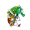
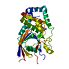
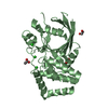
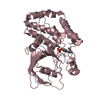
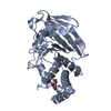
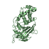
| Title | Human receptor protein tyrosine phosphatase gamma, domain 1, in complex with 2-[(3,4-dichlorobenzyl)sulfanyl]-4-{[3-({N-[2-(methylamino)ethyl]glycyl}amino)phenyl]ethynyl}benzoic acid | ||||||
 Components Components | Receptor-type tyrosine-protein phosphatase gamma | ||||||
 Keywords Keywords | HYDROLASE/HYDROLASE INHIBITOR / TYROSINE RECEPTOR PHOSPHATASE / TWISTED MIXED BETA-SHEETS FLANKED BY {ALPHA}-HELICES / HYDROLASE-HYDROLASE INHIBITOR complex | ||||||
| Function / homology |  Function and homology information Function and homology informationnegative regulation of epithelial cell migration /  transmembrane receptor protein tyrosine phosphatase activity / dephosphorylation / protein-tyrosine-phosphatase / protein tyrosine phosphatase activity / cell surface receptor protein tyrosine kinase signaling pathway / negative regulation of neuron projection development / extracellular exosome / identical protein binding / plasma membrane transmembrane receptor protein tyrosine phosphatase activity / dephosphorylation / protein-tyrosine-phosphatase / protein tyrosine phosphatase activity / cell surface receptor protein tyrosine kinase signaling pathway / negative regulation of neuron projection development / extracellular exosome / identical protein binding / plasma membraneSimilarity search - Function | ||||||
| Biological species |  Homo sapiens (human) Homo sapiens (human) | ||||||
| Method | X-RAY DIFFRACTION / MOLECULAR REPLACEMENT / Resolution: 2.4 Å | ||||||
 Authors Authors | Sheriff, S. | ||||||
 Citation Citation | Journal: J.Med.Chem. / Year: 2011 Title: Small molecule receptor protein tyrosine phosphatase [gamma](RPTP[gamma]) ligands that inhibit phosphatase activity via perturbation of the tryptophan-proline-aspartate (WPD) loop Authors: Sheriff, S. / Beno, B.R. / Zhai, W. / Kostich, W.A. / McDonnell, P.A. / Kish, K. / Goldfarb, V. / Gao, M. / Kiefer, S.E. / Yanchunas, J. / Huang, Y. / Shi, S. / Zhu, S. / Dzierba, C. / ...Authors: Sheriff, S. / Beno, B.R. / Zhai, W. / Kostich, W.A. / McDonnell, P.A. / Kish, K. / Goldfarb, V. / Gao, M. / Kiefer, S.E. / Yanchunas, J. / Huang, Y. / Shi, S. / Zhu, S. / Dzierba, C. / Bronson, J. / Macor, J.E. / Appiah, K.K. / Westphal, R.S. / O'Connell, J. / Gerritz, S.W. #1: Journal: To be PublishedTitle: Cloning, purification, crystallization and preliminary X-ray analysis of the catalytic domain of human receptor-like protein Tyrosine Phosphatase g in three different crystal forms Authors: Kish, K. / McDonnell, P.A. / Goldfarb, V. / Gao, M. / Metzler, W.J. / Langley, D.R. / Bryson, J.W. / Kiefer, S.E. / Kostich, W.A. / Carpenter, B. / Westphal, R.S. / Sheriff, S. | ||||||
| History |
|
- Structure visualization
| Structure viewer | Molecule: MolmilJmol/JSmol |
|---|
- Downloads & links
-Download
| PDBx/mmCIF format | 3qcm.cif.gz | 133.7 KB | Display | PDBx/mmCIF format |
|---|---|---|---|---|
| PDB format | pdb3qcm.ent.gz | 103.4 KB | Display | PDB format |
| PDBx/mmJSON format | 3qcm.json.gz | Tree view | PDBx/mmJSON format | |
| Others |  Other downloads Other downloads |
-Validation report
| Arichive directory | https://data.pdbj.org/pub/pdb/validation_reports/qc/3qcmftp://data.pdbj.org/pub/pdb/validation_reports/qc/3qcm | HTTPS FTP |
|---|
-Related structure data
| Related structure data |  3qcbC 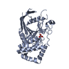 3qccC  3qcdSC  3qceC  3qcfC  3qcgC  3qchC  3qciC  3qcjC  3qckC  3qclC  3qcnC C: citing same article ( S: Starting model for refinement |
|---|---|
| Similar structure data |


-Links
 PDBj
PDBj


- Assembly
| Deposited unit | 
| ||||||||
|---|---|---|---|---|---|---|---|---|---|
| 1 | 
| ||||||||
| 2 | 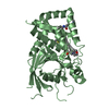
| ||||||||
| Unit cell |
|
-Components
| #1: Protein | Mass: 35735.621 Da / Num. of mol.: 2 / Mutation: V948I, S970T Source method: isolated from a genetically manipulated source Source: (gene. exp.) Homo sapiens (human) / Gene: PTPG, PTPRG / Plasmid: pET28 / Production host:  Escherichia coli (E. coli) / Strain (production host): Rosetta(DE3) / References: UniProt: P23470, protein-tyrosine-phosphatase Escherichia coli (E. coli) / Strain (production host): Rosetta(DE3) / References: UniProt: P23470, protein-tyrosine-phosphatase#2: Chemical |   Mass: 542.477 Da / Num. of mol.: 2 / Source method: obtained synthetically / Formula: C27H25Cl2N3O3S Mass: 542.477 Da / Num. of mol.: 2 / Source method: obtained synthetically / Formula: C27H25Cl2N3O3S#3: Chemical | MES (buffer)  Mass: 195.237 Da / Num. of mol.: 2 / Source method: obtained synthetically / Formula: C6H13NO4S / Comment: pH buffer*YM Mass: 195.237 Da / Num. of mol.: 2 / Source method: obtained synthetically / Formula: C6H13NO4S / Comment: pH buffer*YM#4: Water | ChemComp-HOH / | Water Mass: 18.015 Da / Num. of mol.: 93 / Source method: isolated from a natural source / Formula: H2O Mass: 18.015 Da / Num. of mol.: 93 / Source method: isolated from a natural source / Formula: H2O |
|---|
-Experimental details
-Experiment
| Experiment | Method: X-RAY DIFFRACTION / Number of used crystals: 1 |
|---|
- Sample preparation
| Crystal | Density Matthews: 2.52 Å3/Da / Density % sol: 51.21 % |
|---|---|
| Crystal grow | Temperature: 277 K / pH: 6.7 / Details: pH 6.7, vapor diffusion, temperature 277K |
-Data collection
| Diffraction | Mean temperature: 100 K |
|---|---|
| Diffraction source | Source: ROTATING ANODE / Type: RIGAKU FR-E / Wavelength: 1.54 |
| Detector | Type: RIGAKU RAXIS IV++ / Detector: IMAGE PLATE / Date: May 23, 2007 / Details: MICROMAX CONFOCAL |
| Radiation | Protocol: SINGLE WAVELENGTH / Monochromatic (M) / Laue (L): M / Scattering type: x-ray |
| Radiation wavelength | Wavelength: 1.54 Å / Relative weight: 1 |
| Reflection | Resolution: 2.4→50 Å / Num. obs: 26953 / % possible obs: 97 % / Observed criterion σ(I): 0 / Redundancy: 3.9 % / Biso Wilson estimate: 51.1 Å2 / Rmerge(I) obs: 0.028 / Net I/σ(I): 43 |
| Reflection shell | Resolution: 2.4→2.49 Å / Redundancy: 3.9 % / Rmerge(I) obs: 0.1 / Mean I/σ(I) obs: 14.6 / % possible all: 93.8 |
- Processing
| Software |
| ||||||||||||||||||||||||||||||||||||||||||||||||||||||||||||||||||||||||||||||||||||||||||||||||||||||||||||||||||
|---|---|---|---|---|---|---|---|---|---|---|---|---|---|---|---|---|---|---|---|---|---|---|---|---|---|---|---|---|---|---|---|---|---|---|---|---|---|---|---|---|---|---|---|---|---|---|---|---|---|---|---|---|---|---|---|---|---|---|---|---|---|---|---|---|---|---|---|---|---|---|---|---|---|---|---|---|---|---|---|---|---|---|---|---|---|---|---|---|---|---|---|---|---|---|---|---|---|---|---|---|---|---|---|---|---|---|---|---|---|---|---|---|---|---|---|
| Refinement | Method to determine structure: MOLECULAR REPLACEMENT Starting model: PDB ENTRY 3QCD Resolution: 2.4→40.52 Å / Cor.coef. Fo:Fc: 0.929 / Cor.coef. Fo:Fc free: 0.905 / Occupancy max: 1 / Occupancy min: 0.5 / SU R Cruickshank DPI: 0.39 / Cross valid method: THROUGHOUT / σ(F): 0 / SU R Blow DPI: 0.36 / SU Rfree Blow DPI: 0.24 / SU Rfree Cruickshank DPI: 0.25
| ||||||||||||||||||||||||||||||||||||||||||||||||||||||||||||||||||||||||||||||||||||||||||||||||||||||||||||||||||
| Displacement parameters | Biso mean: 47.07 Å2
| ||||||||||||||||||||||||||||||||||||||||||||||||||||||||||||||||||||||||||||||||||||||||||||||||||||||||||||||||||
| Refine analyze | Luzzati coordinate error obs: 0.31 Å | ||||||||||||||||||||||||||||||||||||||||||||||||||||||||||||||||||||||||||||||||||||||||||||||||||||||||||||||||||
| Refinement step | Cycle: LAST / Resolution: 2.4→40.52 Å
| ||||||||||||||||||||||||||||||||||||||||||||||||||||||||||||||||||||||||||||||||||||||||||||||||||||||||||||||||||
| Refine LS restraints |
| ||||||||||||||||||||||||||||||||||||||||||||||||||||||||||||||||||||||||||||||||||||||||||||||||||||||||||||||||||
| LS refinement shell | Resolution: 2.4→2.49 Å / Total num. of bins used: 14
|
