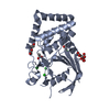
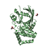
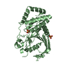
Entry Database : PDB / ID : 5awxTitle Crystal structure of Human PTPRZ D1 domain Receptor-type tyrosine-protein phosphatase zeta Keywords / Function / homology Function Domain/homology Component
/ / / / / / / / / / / / / / / / / / / / / / / / / / / / / / / / / / / / / / / / / / / / / / / / / / / / / / / / / / / / / / / / Biological species Homo sapiens (human)Method / / Resolution : 1.86 Å Authors Sugawara, H. Journal : Sci Rep / Year : 2016Title : Small-molecule inhibition of PTPRZ reduces tumor growth in a rat model of glioblastomaAuthors : Fujikawa, A. / Nagahira, A. / Sugawara, H. / Ishii, K. / Imajo, S. / Matsumoto, M. / Kuboyama, K. / Suzuki, R. / Tanga, N. / Noda, M. / Uchiyama, S. / Tomoo, T. / Ogata, A. / Masumura, M. / Noda, M. History Deposition Jul 10, 2015 Deposition site / Processing site Revision 1.0 Feb 17, 2016 Provider / Type Revision 1.1 Mar 9, 2016 Group Revision 1.2 Nov 8, 2023 Group Data collection / Database references ... Data collection / Database references / Derived calculations / Refinement description Category chem_comp_atom / chem_comp_bond ... chem_comp_atom / chem_comp_bond / database_2 / pdbx_initial_refinement_model / pdbx_struct_oper_list Item / _database_2.pdbx_database_accession / _pdbx_struct_oper_list.symmetry_operation
Show all Show less
 Movie
Movie Controller


 Open data
 Basic information
 Components
 Keywords
 HYDROLASE /
  Function and homology information
 Authors
 Citation
 Structure visualization
 Downloads & links
 Other downloads


 PDBj
PDBj


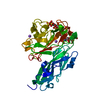
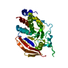
 Assembly
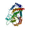


 Mass: 79.904 Da / Num. of mol.: 4 / Source method: obtained synthetically / Formula: Br
 Mass: 18.015 Da / Num. of mol.: 247 / Source method: isolated from a natural source / Formula: H2O
 Sample preparation
 Processing