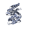
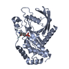
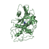
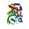
[English] 日本語
Yorodumi
- PDB-5h08: Human PTPRZ D1 domain complexed with NAZ2329 -

+
Open data


ID or keywords:

Loading...

-
Basic information

Entry
Database: PDB / ID: 5h08
TitleHuman PTPRZ D1 domain complexed with NAZ2329
ComponentsReceptor-type tyrosine-protein phosphatase zeta
KeywordsHYDROLASE/HYDROLASE INHIBITOR / Protein Tyrosine Phosphatase / HYDROLASE / HYDROLASE-HYDROLASE INHIBITOR complex
Function / homology
Function and homology information


perineuronal net / regulation of oligodendrocyte progenitor proliferation / transmembrane receptor protein tyrosine phosphatase activity / regulation of myelination / Other interleukin signaling / positive regulation of oligodendrocyte differentiation / Signaling by ALK / oligodendrocyte differentiation / peptidyl-tyrosine dephosphorylation / hematopoietic progenitor cell differentiation ...perineuronal net / regulation of oligodendrocyte progenitor proliferation / transmembrane receptor protein tyrosine phosphatase activity / regulation of myelination / Other interleukin signaling / positive regulation of oligodendrocyte differentiation / Signaling by ALK / oligodendrocyte differentiation / peptidyl-tyrosine dephosphorylation / hematopoietic progenitor cell differentiation / axonogenesis / protein dephosphorylation / protein-tyrosine-phosphatase / central nervous system development / protein tyrosine phosphatase activity / integrin binding / negative regulation of neuron apoptotic process / learning or memory / synapse / extracellular region / plasma membrane
Similarity search - Function
Receptor-type tyrosine-protein phosphatase, carbonic anhydrase domain / Protein tyrosine phosphatase superfamily / Protein-Tyrosine Phosphatase; Chain A / Protein tyrosine phosphatase, catalytic domain / PTP type protein phosphatase domain profile. / Protein-tyrosine phosphatase / Tyrosine-specific protein phosphatase, PTPase domain / Protein-tyrosine phosphatase, catalytic / Protein tyrosine phosphatase, catalytic domain motif / Tyrosine specific protein phosphatases active site. ...Receptor-type tyrosine-protein phosphatase, carbonic anhydrase domain / Protein tyrosine phosphatase superfamily / Protein-Tyrosine Phosphatase; Chain A / Protein tyrosine phosphatase, catalytic domain / PTP type protein phosphatase domain profile. / Protein-tyrosine phosphatase / Tyrosine-specific protein phosphatase, PTPase domain / Protein-tyrosine phosphatase, catalytic / Protein tyrosine phosphatase, catalytic domain motif / Tyrosine specific protein phosphatases active site. / Protein-tyrosine phosphatase, active site / Tyrosine-specific protein phosphatases domain / Tyrosine specific protein phosphatases domain profile. / Protein-tyrosine phosphatase-like / Fibronectin type III domain / Alpha carbonic anhydrase domain / Alpha carbonic anhydrase domain superfamily / Eukaryotic-type carbonic anhydrase / Alpha-carbonic anhydrases profile. / Eukaryotic-type carbonic anhydrase / Fibronectin type 3 domain / Fibronectin type-III domain profile. / Fibronectin type III / Fibronectin type III superfamily / Immunoglobulin-like fold / Alpha-Beta Complex / Alpha Beta
Similarity search - Domain/homology
Chem-7WL / Receptor-type tyrosine-protein phosphatase zeta
Similarity search - Component
Biological speciesHomo sapiens (human)
MethodX-RAY DIFFRACTION / MOLECULAR REPLACEMENT / Resolution: 2.53 Å
AuthorsSugawara, H.
Funding support Japan, 1items
OrganizationGrant numberCountry
Japan Agency for Medical Research and development15im0110409b0104 Japan
Citation
Journal: Sci Rep / Year: 2017
Title: Targeting PTPRZ inhibits stem cell-like properties and tumorigenicity in glioblastoma cells
Authors: Fujikawa, A. / Sugawara, H. / Tanaka, T. / Matsumoto, M. / Kuboyama, K. / Suzuki, R. / Tanga, N. / Ogata, A. / Masumura, M. / Noda, M.
#1: Journal: Scientific Reports / Year: 2016
Title: Small-molecule inhibition of PTPRZ suppresses tumor growth in a rat model of glioblastoma.
Authors: Fujikawa, A. / Nagahira, A. / Sugawara, H. / Ishii, K. / Imajo, S. / Matsumoto, M. / Kuboyama, K. / Suzuki, R. / Tanga, N. / Noda, M. / Uchiyama, S. / Tomoo, T. / Ogata, A. / Masumura, M. / Noda, M.
History
DepositionOct 4, 2016Deposition site: PDBJ / Processing site: PDBJ
Revision 1.0Jul 26, 2017Provider: repository / Type: Initial release
Revision 1.1Aug 16, 2017Group: Database references / Category: citation
Item: _citation.journal_abbrev / _citation.journal_volume ..._citation.journal_abbrev / _citation.journal_volume / _citation.page_first / _citation.page_last / _citation.pdbx_database_id_DOI / _citation.pdbx_database_id_PubMed
Revision 1.2Nov 8, 2023Group: Data collection / Database references / Refinement description
Category: chem_comp_atom / chem_comp_bond ...chem_comp_atom / chem_comp_bond / database_2 / pdbx_initial_refinement_model
Item: _database_2.pdbx_DOI / _database_2.pdbx_database_accession

-
Structure visualization

Structure viewerMolecule:
MolmilJmol/JSmol

Downloads & links

-
Assembly

Deposited unit
A: Receptor-type tyrosine-protein phosphatase zeta
hetero molecules


Theoretical massNumber of molelcules
Total (without water)34,4562
Polymers33,9541
Non-polymers5021
Water25214
1


  • Idetical with deposited unit
  • defined by author
TypeNameSymmetry operationNumber
identity operation1_555x,y,z1
Buried area0 Å2
ΔGint0 kcal/mol
Surface area13350 Å2
Unit cell
Length a, b, c (Å)53.776, 72.306, 90.796
Angle α, β, γ (deg.)90.00, 90.00, 90.00
Int Tables number19
Space group name H-MP212121
DetailsTHIS PROTEIN IS ACTIVE MONOMER, HOWEVER IT DIMERIZES BY

-
Components

#1: Protein Receptor-type tyrosine-protein phosphatase zeta / R-PTP-zeta / Protein-tyrosine phosphatase receptor type Z polypeptide 1 / Protein-tyrosine ...R-PTP-zeta / Protein-tyrosine phosphatase receptor type Z polypeptide 1 / Protein-tyrosine phosphatase receptor type Z polypeptide 2 / R-PTP-zeta-2


Mass: 33954.402 Da / Num. of mol.: 1 / Fragment: UNP residues 1698-1993
Source method: isolated from a genetically manipulated source
Source: (gene. exp.) Homo sapiens (human) / Gene: PTPRZ1, HTPZP2, PTPRZ, PTPRZ2, PTPZ / Production host: Bombyx mori (domestic silkworm) / References: UniProt: P23471, protein-tyrosine-phosphatase
#2: Chemical ChemComp-7WL / 3-{[2-Ethoxy-5-(trifluoromethyl)benzyl]sulfanyl}-N-(phenylsulfonyl)thiophene-2-carboxamide


Mass: 501.562 Da / Num. of mol.: 1 / Source method: obtained synthetically / Formula: C21H18F3NO4S3
#3: Water ChemComp-HOH / water / Water


Mass: 18.015 Da / Num. of mol.: 14 / Source method: isolated from a natural source / Formula: H2O
Sequence detailsTHIS SEQUENCE CORRESPONDS TO THE ISOFORM 2 IN UNP P23471.

-
Experimental details

-
Experiment

ExperimentMethod: X-RAY DIFFRACTION / Number of used crystals: 1

-
Sample preparation

CrystalDensity Matthews: 2.6 Å3/Da / Density % sol: 52.68 %
Crystal growTemperature: 277 K / Method: vapor diffusion, sitting drop / pH: 8.5
Details: 10%(w/v) PEG8000, 20%(v/v) ethylene glycol, 30mM sodium fluoride, 30mM sodium bromide, 30mM sodium iodide. NAZ2329 was soaked into the crystal.

-
Data collection

DiffractionMean temperature: 100 K
Diffraction sourceSource: ROTATING ANODE / Type: RIGAKU MICROMAX-007 / Wavelength: 1.5418 Å
DetectorType: RIGAKU / Detector: IMAGE PLATE / Date: Apr 9, 2015
RadiationProtocol: SINGLE WAVELENGTH / Monochromatic (M) / Laue (L): M / Scattering type: x-ray
Radiation wavelengthWavelength: 1.5418 Å / Relative weight: 1
ReflectionResolution: 2.53→100 Å / Num. obs: 10798 / % possible obs: 87.2 % / Redundancy: 4.5 % / Rmerge(I) obs: 0.03 / Net I/σ(I): 30.6

-
Processing

Software
NameVersionClassification
REFMAC5.8.0158refinement
HKL-2000data reduction
HKL-2000data scaling
AMoREphasing
RefinementMethod to determine structure: MOLECULAR REPLACEMENT
Starting model: 5AWX
Resolution: 2.53→45.4 Å / Cor.coef. Fo:Fc: 0.917 / Cor.coef. Fo:Fc free: 0.852 / SU B: 25.429 / SU ML: 0.262 / Cross valid method: THROUGHOUT / ESU R: 0.219 / ESU R Free: 0.082 / Stereochemistry target values: MAXIMUM LIKELIHOOD / Details: HYDROGENS HAVE BEEN ADDED IN THE RIDING POSITIONS
RfactorNum. reflection% reflectionSelection details
Rfree0.29689 536 5.1 %RANDOM
Rwork0.21029 ---
obs0.21476 9992 84.97 %-
Solvent computationIon probe radii: 0.8 Å / Shrinkage radii: 0.8 Å / VDW probe radii: 1.2 Å / Solvent model: MASK
Displacement parametersBiso mean: 39.25 Å2
Baniso -1Baniso -2Baniso -3
1--29.55 Å20 Å20 Å2
2--11.65 Å20 Å2
3---17.9 Å2
Refinement stepCycle: 1 / Resolution: 2.53→45.4 Å
ProteinNucleic acidLigandSolventTotal
Num. atoms2276 0 32 14 2322
Refine LS restraints
Refine-IDTypeDev idealDev ideal targetNumber
X-RAY DIFFRACTIONr_bond_refined_d0.0170.0192367
X-RAY DIFFRACTIONr_bond_other_d0.0020.022129
X-RAY DIFFRACTIONr_angle_refined_deg1.8941.9443219
X-RAY DIFFRACTIONr_angle_other_deg1.08334926
X-RAY DIFFRACTIONr_dihedral_angle_1_deg7.8775280
X-RAY DIFFRACTIONr_dihedral_angle_2_deg37.66723.793116
X-RAY DIFFRACTIONr_dihedral_angle_3_deg18.40715384
X-RAY DIFFRACTIONr_dihedral_angle_4_deg18.9881514
X-RAY DIFFRACTIONr_chiral_restr0.1020.2350
X-RAY DIFFRACTIONr_gen_planes_refined0.0080.0212625
X-RAY DIFFRACTIONr_gen_planes_other0.0020.02505
X-RAY DIFFRACTIONr_nbd_refined
X-RAY DIFFRACTIONr_nbd_other
X-RAY DIFFRACTIONr_nbtor_refined
X-RAY DIFFRACTIONr_nbtor_other
X-RAY DIFFRACTIONr_xyhbond_nbd_refined
X-RAY DIFFRACTIONr_xyhbond_nbd_other
X-RAY DIFFRACTIONr_metal_ion_refined
X-RAY DIFFRACTIONr_metal_ion_other
X-RAY DIFFRACTIONr_symmetry_vdw_refined
X-RAY DIFFRACTIONr_symmetry_vdw_other
X-RAY DIFFRACTIONr_symmetry_hbond_refined
X-RAY DIFFRACTIONr_symmetry_hbond_other
X-RAY DIFFRACTIONr_symmetry_metal_ion_refined
X-RAY DIFFRACTIONr_symmetry_metal_ion_other
X-RAY DIFFRACTIONr_mcbond_it0.7091.6111126
X-RAY DIFFRACTIONr_mcbond_other0.7091.6111125
X-RAY DIFFRACTIONr_mcangle_it1.2612.4151404
X-RAY DIFFRACTIONr_mcangle_other1.2612.4161405
X-RAY DIFFRACTIONr_scbond_it0.5281.6241241
X-RAY DIFFRACTIONr_scbond_other0.5291.6251242
X-RAY DIFFRACTIONr_scangle_it
X-RAY DIFFRACTIONr_scangle_other0.9552.4271816
X-RAY DIFFRACTIONr_long_range_B_refined2.37718.4512679
X-RAY DIFFRACTIONr_long_range_B_other2.37618.4522680
X-RAY DIFFRACTIONr_rigid_bond_restr
X-RAY DIFFRACTIONr_sphericity_free
X-RAY DIFFRACTIONr_sphericity_bonded
LS refinement shellResolution: 2.526→2.592 Å / Total num. of bins used: 20
RfactorNum. reflection% reflection
Rfree0.395 17 -
Rwork0.293 373 -
obs--42.9 %
Refinement TLS params.Method: refined / Origin x: 19.9262 Å / Origin y: 0.4235 Å / Origin z: 44.6027 Å
111213212223313233
T0.2331 Å20.0147 Å2-0.0195 Å2-0.039 Å2-0.0075 Å2--0.0354 Å2
L3.3654 °2-0.4957 °20.216 °2-3.2108 °20.8278 °2--2.2589 °2
S0.0244 Å °-0.0037 Å °-0.3331 Å °0.0552 Å °0.0697 Å °0.0245 Å °0.3861 Å °0.2686 Å °-0.0941 Å °

+
About Yorodumi

-
News

-
Feb 9, 2022. New format data for meta-information of EMDB entries

New format data for meta-information of EMDB entries

  • Version 3 of the EMDB header file is now the official format.
  • The previous official version 1.9 will be removed from the archive.

Related info.:EMDB header

External links:wwPDB to switch to version 3 of the EMDB data model

-
Aug 12, 2020. Covid-19 info

Covid-19 info

URL: https://pdbj.org/emnavi/covid19.php

New page: Covid-19 featured information page in EM Navigator.

Related info.:Covid-19 info / Mar 5, 2020. Novel coronavirus structure data

+
Mar 5, 2020. Novel coronavirus structure data

Novel coronavirus structure data

Related info.:Yorodumi Speices / Aug 12, 2020. Covid-19 info

External links:COVID-19 featured content - PDBj / Molecule of the Month (242):Coronavirus Proteases

+
Jan 31, 2019. EMDB accession codes are about to change! (news from PDBe EMDB page)

EMDB accession codes are about to change! (news from PDBe EMDB page)

  • The allocation of 4 digits for EMDB accession codes will soon come to an end. Whilst these codes will remain in use, new EMDB accession codes will include an additional digit and will expand incrementally as the available range of codes is exhausted. The current 4-digit format prefixed with “EMD-” (i.e. EMD-XXXX) will advance to a 5-digit format (i.e. EMD-XXXXX), and so on. It is currently estimated that the 4-digit codes will be depleted around Spring 2019, at which point the 5-digit format will come into force.
  • The EM Navigator/Yorodumi systems omit the EMD- prefix.

Related info.:Q: What is EMD? / ID/Accession-code notation in Yorodumi/EM Navigator

External links:EMDB Accession Codes are Changing Soon! / Contact to PDBj

+
Jul 12, 2017. Major update of PDB

Major update of PDB

  • wwPDB released updated PDB data conforming to the new PDBx/mmCIF dictionary.
  • This is a major update changing the version number from 4 to 5, and with Remediation, in which all the entries are updated.
  • In this update, many items about electron microscopy experimental information are reorganized (e.g. em_software).
  • Now, EM Navigator and Yorodumi are based on the updated data.

External links:wwPDB Remediation / Enriched Model Files Conforming to OneDep Data Standards Now Available in the PDB FTP Archive

-
Yorodumi

Thousand views of thousand structures

  • Yorodumi is a browser for structure data from EMDB, PDB, SASBDB, etc.
  • This page is also the successor to EM Navigator detail page, and also detail information page/front-end page for Omokage search.
  • The word "yorodu" (or yorozu) is an old Japanese word meaning "ten thousand". "mi" (miru) is to see.

Related info.:EMDB / PDB / SASBDB / Comparison of 3 databanks / Yorodumi Search / Aug 31, 2016. New EM Navigator & Yorodumi / Yorodumi Papers / Jmol/JSmol / Function and homology information / Changes in new EM Navigator and Yorodumi

Read more