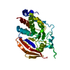
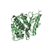
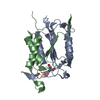
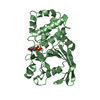
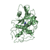
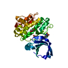
BIOMOLECULE: 1 THIS ENTRY CONTAINS THE CRYSTALLOGRAPHIC ASYMMETRIC UNIT WHICH CONSISTS OF 2 CHAINS. ... BIOMOLECULE: 1 THIS ENTRY CONTAINS THE CRYSTALLOGRAPHIC ASYMMETRIC UNIT WHICH CONSISTS OF 2 CHAINS. SEE REMARK 350 FOR INFORMATION ON GENERATING THE BIOLOGICAL MOLECULE(S). SIZE EXCLUSION CHROMATOGRAPHY SUGGESTS THE ASSIGNMENT OF A DIMER AS THE SIGNIFICANT OLIGOMERIZATION STATE.
Remark 999
SEQUENCE THE CONSTRUCT WAS EXPRESSED WITH A PURIFICATION TAG MGSDKIHHHHHHENLYFQG. THE TAG WAS ... SEQUENCE THE CONSTRUCT WAS EXPRESSED WITH A PURIFICATION TAG MGSDKIHHHHHHENLYFQG. THE TAG WAS PARTIALLY REMOVED WITH TEV PROTEASE LEAVING ONLY A GLYCINE FOLLOWED BY THE TARGET SEQUENCE.
Monochromator: Single crystal Si(111) bent (horizontal focusing) Protocol: MAD / Monochromatic (M) / Laue (L): M / Scattering type: x-ray
Radiation wavelength
ID
Wavelength (Å)
Relative weight
1
0.91837
1
2
0.97917
1
3
0.97891
1
Reflection
Resolution: 1.5→29.761 Å / Num. obs: 71427 / % possible obs: 99.6 % / Observed criterion σ(I): -3 / Biso Wilson estimate: 24.94 Å2 / Rmerge(I) obs: 0.04 / Net I/σ(I): 18.13
Reflection shell
Resolution (Å)
Rmerge(I) obs
Mean I/σ(I) obs
Num. measured obs
Num. unique obs
Diffraction-ID
% possible all
1.5-1.55
0.517
2.5
46448
12464
1
97.4
1.55-1.62
0.391
3.3
57472
15201
1
100
1.62-1.69
0.297
4.4
48686
12823
1
100
1.69-1.78
0.209
6.2
52186
13705
1
100
1.78-1.89
0.139
9.1
50978
13337
1
99.9
1.89-2.04
0.085
14.3
53269
13885
1
100
2.04-2.24
0.054
21.5
50781
13184
1
100
2.24-2.56
0.04
28.2
52007
13475
1
99.9
2.56-3.23
0.027
39.2
52846
13677
1
99.9
3.23-29.761
0.021
52.8
51585
13480
1
98.8
-
Phasing
Phasing
Method: MAD
-
Processing
Software
Name
Version
Classification
NB
REFMAC
5.2.0019
refinement
PHENIX
refinement
SHELX
phasing
MolProbity
3beta29
modelbuilding
XSCALE
datascaling
PDB_EXTRACT
3
dataextraction
MAR345
CCD
datacollection
XDS
datareduction
SHELXD
phasing
autoSHARP
phasing
Refinement
Method to determine structure: MAD / Resolution: 1.5→29.761 Å / Cor.coef. Fo:Fc: 0.976 / Cor.coef. Fo:Fc free: 0.97 / SU B: 1.698 / SU ML: 0.029 / Cross valid method: THROUGHOUT / σ(F): 0 / ESU R: 0.052 / ESU R Free: 0.049 Stereochemistry target values: MAXIMUM LIKELIHOOD WITH PHASES Details: 1. HYDROGENS HAVE BEEN ADDED IN THE RIDING POSITIONS. 2. A MET-INHIBITION PROTOCOL WAS USED FOR SELENOMETHIONINE INCORPORATION DURING PROTEIN EXPRESSION. THE OCCUPANCY OF THE SE ATOMS IN THE ...Details: 1. HYDROGENS HAVE BEEN ADDED IN THE RIDING POSITIONS. 2. A MET-INHIBITION PROTOCOL WAS USED FOR SELENOMETHIONINE INCORPORATION DURING PROTEIN EXPRESSION. THE OCCUPANCY OF THE SE ATOMS IN THE MSE RESIDUES WAS REDUCED TO 0.75 FOR THE REDUCED SCATTERING POWER DUE TO PARTIAL S-MET INCORPORATION. 3. ETHYLENE GLYCOL IS MODELED BASED ON CRYO CONDITIONS. 4. THE BENZOIC ACID (BEZ) MOLECULE IS ASSIGNED BASED ON THE ELECTRON DENSITY AND ITS INTERACTION WITH THE PROTEIN. IT COULD BE SOME RELATED COMPOUND WITH A SIMILAR STRUCTURE. 5. RESIDUES A0 AND B0-1 ARE DISORDERED AND WERE NOT MODELED.
Rfactor
Num. reflection
% reflection
Selection details
Rfree
0.159
3613
5.1 %
RANDOM
Rwork
0.135
-
-
-
obs
0.136
71338
99.85 %
-
Solvent computation
Ion probe radii: 0.8 Å / Shrinkage radii: 0.8 Å / VDW probe radii: 1.2 Å / Solvent model: MASK
Displacement parameters
Biso mean: 15.81 Å2
Baniso -1
Baniso -2
Baniso -3
1-
-0.03 Å2
0 Å2
0 Å2
2-
-
-0.03 Å2
0 Å2
3-
-
-
0.05 Å2
Refinement step
Cycle: LAST / Resolution: 1.5→29.761 Å
Protein
Nucleic acid
Ligand
Solvent
Total
Num. atoms
2162
0
54
431
2647
Refine LS restraints
Refine-ID
Type
Dev ideal
Dev ideal target
Number
X-RAY DIFFRACTION
r_bond_refined_d
0.017
0.021
2353
X-RAY DIFFRACTION
r_bond_other_d
0.004
0.02
1561
X-RAY DIFFRACTION
r_angle_refined_deg
1.538
1.966
3209
X-RAY DIFFRACTION
r_angle_other_deg
0.97
3
3845
X-RAY DIFFRACTION
r_dihedral_angle_1_deg
5.724
5
314
X-RAY DIFFRACTION
r_dihedral_angle_2_deg
37.795
25.14
107
X-RAY DIFFRACTION
r_dihedral_angle_3_deg
11.056
15
382
X-RAY DIFFRACTION
r_dihedral_angle_4_deg
13.592
15
10
X-RAY DIFFRACTION
r_chiral_restr
0.091
0.2
365
X-RAY DIFFRACTION
r_gen_planes_refined
0.007
0.02
2662
X-RAY DIFFRACTION
r_gen_planes_other
0.001
0.02
440
X-RAY DIFFRACTION
r_nbd_refined
0.222
0.2
428
X-RAY DIFFRACTION
r_nbd_other
0.196
0.2
1655
X-RAY DIFFRACTION
r_nbtor_refined
0.172
0.2
1134
X-RAY DIFFRACTION
r_nbtor_other
0.089
0.2
1142
X-RAY DIFFRACTION
r_xyhbond_nbd_refined
0.176
0.2
249
X-RAY DIFFRACTION
r_xyhbond_nbd_other
0.146
0.2
1
X-RAY DIFFRACTION
r_symmetry_vdw_refined
0.226
0.2
10
X-RAY DIFFRACTION
r_symmetry_vdw_other
0.272
0.2
40
X-RAY DIFFRACTION
r_symmetry_hbond_refined
0.208
0.2
24
X-RAY DIFFRACTION
r_mcbond_it
2.924
3
1537
X-RAY DIFFRACTION
r_mcbond_other
2.043
3
589
X-RAY DIFFRACTION
r_mcangle_it
3.669
5
2378
X-RAY DIFFRACTION
r_scbond_it
4.858
8
942
X-RAY DIFFRACTION
r_scangle_it
6.453
11
814
X-RAY DIFFRACTION
r_rigid_bond_restr
2.55
3
4286
X-RAY DIFFRACTION
r_sphericity_free
8.828
3
431
X-RAY DIFFRACTION
r_sphericity_bonded
5.326
3
3859
LS refinement shell
Resolution: 1.5→1.539 Å / Total num. of bins used: 20
Rfactor
Num. reflection
% reflection
Rfree
0.245
230
-
Rwork
0.16
4922
-
obs
-
5152
98.81 %
+
About Yorodumi
-
News
-
Feb 9, 2022. New format data for meta-information of EMDB entries
New format data for meta-information of EMDB entries
Version 3 of the EMDB header file is now the official format.
The previous official version 1.9 will be removed from the archive.
In the structure databanks used in Yorodumi, some data are registered as the other names, "COVID-19 virus" and "2019-nCoV". Here are the details of the virus and the list of structure data.
Jan 31, 2019. EMDB accession codes are about to change! (news from PDBe EMDB page)
EMDB accession codes are about to change! (news from PDBe EMDB page)
The allocation of 4 digits for EMDB accession codes will soon come to an end. Whilst these codes will remain in use, new EMDB accession codes will include an additional digit and will expand incrementally as the available range of codes is exhausted. The current 4-digit format prefixed with “EMD-” (i.e. EMD-XXXX) will advance to a 5-digit format (i.e. EMD-XXXXX), and so on. It is currently estimated that the 4-digit codes will be depleted around Spring 2019, at which point the 5-digit format will come into force.
The EM Navigator/Yorodumi systems omit the EMD- prefix.
Related info.:Q: What is EMD? / ID/Accession-code notation in Yorodumi/EM Navigator
Yorodumi is a browser for structure data from EMDB, PDB, SASBDB, etc.
This page is also the successor to EM Navigator detail page, and also detail information page/front-end page for Omokage search.
The word "yorodu" (or yorozu) is an old Japanese word meaning "ten thousand". "mi" (miru) is to see.
Related info.:EMDB / PDB / SASBDB / Comparison of 3 databanks / Yorodumi Search / Aug 31, 2016. New EM Navigator & Yorodumi / Yorodumi Papers / Jmol/JSmol / Function and homology information / Changes in new EM Navigator and Yorodumi
 Movie
Movie Controller

 Yorodumi
 Open data
 Basic information
 Components
 Keywords
 Function and homology information
 Authors
 Citation
 Structure visualization
 Downloads & links
 Other downloads


 PDBj
PDBj Assembly

 Mass: 122.121 Da / Num. of mol.: 2 / Source method: obtained synthetically / Formula: C7H6O2
 Mass: 62.068 Da / Num. of mol.: 9 / Source method: obtained synthetically / Formula: C2H6O2
 Mass: 18.015 Da / Num. of mol.: 431 / Source method: isolated from a natural source / Formula: H2O
 Sample preparation
 / Beamline: BL11-1 / Wavelength: 0.91837, 0.97917, 0.97891
 Processing