 Movie
Movie Controller

[English] 日本語
 Yorodumi
- PDB-1p2x: CRYSTAL STRUCTURE OF THE CALPONIN-HOMOLOGY DOMAIN OF RNG2 FROM SC... -
+ Open data

- Basic information
| Entry | Database: PDB / ID: 1p2x | ||||||
|---|---|---|---|---|---|---|---|
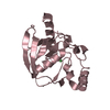
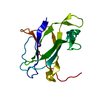
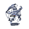
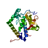
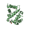
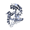
| Title | CRYSTAL STRUCTURE OF THE CALPONIN-HOMOLOGY DOMAIN OF RNG2 FROM SCHIZOSACCHAROMYCES POMBE | ||||||
 Components Components | Ras GTPase-activating-like protein | ||||||
 Keywords Keywords |  PROTEIN BINDING / 4 HELICES / BUNDLE PROTEIN BINDING / 4 HELICES / BUNDLE | ||||||
| Function / homology |  Function and homology information Function and homology informationactomyosin contractile ring assembly actin filament bundle convergence / RHOV GTPase cycle / RHOA GTPase cycle / RHO GTPases activate IQGAPs / medial cortical node / mitotic actomyosin contractile ring, proximal layer / mitotic actomyosin contractile ring assembly actin filament organization / RHOQ GTPase cycle / RHOU GTPase cycle / Neutrophil degranulation ...actomyosin contractile ring assembly actin filament bundle convergence / RHOV GTPase cycle / RHOA GTPase cycle / RHO GTPases activate IQGAPs / medial cortical node / mitotic actomyosin contractile ring, proximal layer / mitotic actomyosin contractile ring assembly actin filament organization / RHOQ GTPase cycle / RHOU GTPase cycle / Neutrophil degranulation / mitotic actomyosin contractile ring assembly / mitotic spindle pole body / mitotic actomyosin contractile ring / mitotic actomyosin contractile ring contraction / cytoskeletal anchor activity / spindle pole body / GTPase activator activity / actin filament binding / nuclear envelope / cell cortex / calmodulin binding / cytoplasmSimilarity search - Function | ||||||
| Biological species |  Schizosaccharomyces pombe (fission yeast) Schizosaccharomyces pombe (fission yeast) | ||||||
| Method | X-RAY DIFFRACTION / SYNCHROTRON / MAD / Resolution: 2.21 Å | ||||||
 Authors Authors | Wang, C.-H. / Balasubramanian, M.K. / Dokland, T. | ||||||
 Citation Citation | Journal: Acta Crystallogr.,Sect.D / Year: 2004 Title: Structure, crystal packing and molecular dynamics of the calponin-homology domain of Schizosaccharomyces pombe Rng2. Authors: Wang, C.H. / Balasubramanian, M.K. / Dokland, T. | ||||||
| History |
|
- Structure visualization
| Structure viewer | Molecule: MolmilJmol/JSmol |
|---|
- Downloads & links
-Download
| PDBx/mmCIF format | 1p2x.cif.gz | 49 KB | Display | PDBx/mmCIF format |
|---|---|---|---|---|
| PDB format | pdb1p2x.ent.gz | 33.9 KB | Display | PDB format |
| PDBx/mmJSON format | 1p2x.json.gz | Tree view | PDBx/mmJSON format | |
| Others |  Other downloads Other downloads |
-Validation report
| Arichive directory | https://data.pdbj.org/pub/pdb/validation_reports/p2/1p2xftp://data.pdbj.org/pub/pdb/validation_reports/p2/1p2x | HTTPS FTP |
|---|
-Related structure data


-Links
 PDBj
PDBj


- Assembly
| Deposited unit | 
| ||||||||
|---|---|---|---|---|---|---|---|---|---|
| 1 |
| ||||||||
| Unit cell |
|
-Components
| #1: Protein | Mass: 18476.211 Da / Num. of mol.: 1 / Fragment: CALPONIN-HOMOLOGY DOMAIN Source method: isolated from a genetically manipulated source Source: (gene. exp.) Schizosaccharomyces pombe (fission yeast)Gene: RNG2 OR SPAC4F8.13C / Plasmid: pGEX-KG / Production host:  Escherichia coli (E. coli) / Strain (production host): BL21-CodonPlus-RIL / References: UniProt: O14188 Escherichia coli (E. coli) / Strain (production host): BL21-CodonPlus-RIL / References: UniProt: O14188 | ||
|---|---|---|---|
| #2: Chemical | ChemComp-BR / Bromide  Mass: 79.904 Da / Num. of mol.: 11 / Source method: obtained synthetically / Formula: Br Mass: 79.904 Da / Num. of mol.: 11 / Source method: obtained synthetically / Formula: Br#3: Water | ChemComp-HOH / | Water Mass: 18.015 Da / Num. of mol.: 157 / Source method: isolated from a natural source / Formula: H2O Mass: 18.015 Da / Num. of mol.: 157 / Source method: isolated from a natural source / Formula: H2O |
-Experimental details
-Experiment
| Experiment | Method: X-RAY DIFFRACTION / Number of used crystals: 1 |
|---|
- Sample preparation
| Crystal | Density Matthews: 2 Å3/Da / Density % sol: 38.07 % |
|---|---|
| Crystal grow | Temperature: 295 K / Method: vapor diffusion, sitting drop / pH: 7 Details: 21% PEG 3000, 0.3M Calcium Acetate, 0.1M Tris pH 7, VAPOR DIFFUSION, SITTING DROP, temperature 295K |
-Data collection
| Diffraction | Mean temperature: 100 K | ||||||||||||
|---|---|---|---|---|---|---|---|---|---|---|---|---|---|
| Diffraction source | Source: SYNCHROTRON / Site: ESRF  / Beamline: BM14 / Wavelength: 0.919087, 0.919338, 0.855057 / Beamline: BM14 / Wavelength: 0.919087, 0.919338, 0.855057 | ||||||||||||
| Detector | Type: MARRESEARCH / Detector: CCD / Date: Sep 15, 2002 / Details: mirrors | ||||||||||||
| Radiation | Monochromator: Si(111) / Protocol: MAD / Monochromatic (M) / Laue (L): M / Scattering type: x-ray | ||||||||||||
| Radiation wavelength |
| ||||||||||||
| Reflection | Resolution: 2.21→22.65 Å / Num. obs: 7532 / % possible obs: 89.6 % / Observed criterion σ(F): 3 / Observed criterion σ(I): 3 / Redundancy: 3.47 % / Biso Wilson estimate: 16.8 Å2 / Rmerge(I) obs: 0.053 / Rsym value: 0.053 / Net I/σ(I): 0.053 | ||||||||||||
| Reflection shell | Resolution: 2.21→2.29 Å / Redundancy: 2.09 % / Rmerge(I) obs: 0.077 / Mean I/σ(I) obs: 9.39 / Num. unique all: 202 / Rsym value: 0.077 / % possible all: 24.8 |
- Processing
| Software |
| |||||||||||||||||||||||||
|---|---|---|---|---|---|---|---|---|---|---|---|---|---|---|---|---|---|---|---|---|---|---|---|---|---|---|
| Refinement | Method to determine structure: MAD / Resolution: 2.21→22.65 Å / Rfactor Rfree error: 0.014 / Data cutoff high absF: 930480.25 / Data cutoff high rms absF: 930480.25 / Data cutoff low absF: 0 / Isotropic thermal model: RESTRAINED / Cross valid method: THROUGHOUT / σ(F): 3 / σ(I): 3 / Stereochemistry target values: Engh & Huber
| |||||||||||||||||||||||||
| Solvent computation | Solvent model: FLAT MODEL / Bsol: 43.4937 Å2 / ksol: 0.346508 e/Å3 | |||||||||||||||||||||||||
| Displacement parameters | Biso mean: 17.6 Å2
| |||||||||||||||||||||||||
| Refine analyze |
| |||||||||||||||||||||||||
| Refinement step | Cycle: LAST / Resolution: 2.21→22.65 Å
| |||||||||||||||||||||||||
| Refine LS restraints |
| |||||||||||||||||||||||||
| LS refinement shell | Resolution: 2.21→2.35 Å / Rfactor Rfree error: 0.043 / Total num. of bins used: 6
| |||||||||||||||||||||||||
| Xplor file |
|
