 Movie
Movie Controller

[English] 日本語
 Yorodumi
- PDB-1yp1: Crystal structure of a non-hemorrhagic fibrin(ogen)olytic metallo... -
+ Open data

- Basic information
| Entry | Database: PDB / ID: 1yp1 | ||||||
|---|---|---|---|---|---|---|---|
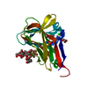
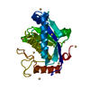
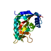
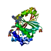
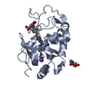
| Title | Crystal structure of a non-hemorrhagic fibrin(ogen)olytic metalloproteinase from venom of Agkistrodon acutus | ||||||
 Components Components |
| ||||||
 Keywords Keywords |  HYDROLASE / FII crystal structure HYDROLASE / FII crystal structure | ||||||
| Function / homology | Collagenase (Catalytic Domain) / Collagenase (Catalytic Domain) / 3-Layer(aba) Sandwich / Alpha Beta Function and homology information Function and homology information | ||||||
| Biological species |  Deinagkistrodon acutus (Chinese moccasin) Deinagkistrodon acutus (Chinese moccasin) | ||||||
| Method | X-RAY DIFFRACTION / MOLECULAR REPLACEMENT / Resolution: 1.9 Å | ||||||
 Authors Authors | Lou, Z. / Hou, J. / Chen, J. / Liang, X. / Qiu, P. / Liu, Y. / Li, M. / Rao, Z. | ||||||
 Citation Citation | Journal: J.Struct.Biol. / Year: 2005 Title: Crystal structure of a non-hemorrhagic fibrin(ogen)olytic metalloproteinase complexed with a novel natural tri-peptide inhibitor from venom of Agkistrodon acutus Authors: Lou, Z. / Hou, J. / Liang, X. / Chen, J. / Qiu, P. / Liu, Y. / Li, M. / Rao, Z. / Yan, G. | ||||||
| History |
|
- Structure visualization
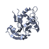
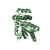
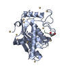
| Structure viewer | Molecule: MolmilJmol/JSmol |
|---|
- Downloads & links
-Download
| PDBx/mmCIF format | 1yp1.cif.gz | 49.8 KB | Display | PDBx/mmCIF format |
|---|---|---|---|---|
| PDB format | pdb1yp1.ent.gz | 38.6 KB | Display | PDB format |
| PDBx/mmJSON format | 1yp1.json.gz | Tree view | PDBx/mmJSON format | |
| Others |  Other downloads Other downloads |
-Validation report
| Arichive directory | https://data.pdbj.org/pub/pdb/validation_reports/yp/1yp1ftp://data.pdbj.org/pub/pdb/validation_reports/yp/1yp1 | HTTPS FTP |
|---|
-Related structure data
| Similar structure data |
|---|

-Links
 PDBj
PDBj- Assembly
| Deposited unit | 
| ||||||||
|---|---|---|---|---|---|---|---|---|---|
| 1 |
| ||||||||
| Unit cell |
|
-Components
| #1: Protein | Mass: 21987.357 Da / Num. of mol.: 1 / Source method: isolated from a natural source / Source: (natural) Deinagkistrodon acutus (Chinese moccasin)References: Hydrolases; Acting on peptide bonds (peptidases); Metalloendopeptidases |
|---|---|
| #2: Protein/peptide | Mass: 374.456 Da / Num. of mol.: 1 / Source method: obtained synthetically / Details: This sequence occurs naturally synthesis |
| #3: Chemical | ChemComp-ZN /   Mass: 65.409 Da / Num. of mol.: 1 / Source method: obtained synthetically / Formula: Zn Mass: 65.409 Da / Num. of mol.: 1 / Source method: obtained synthetically / Formula: Zn |
| #4: Water | ChemComp-HOH / Water Mass: 18.015 Da / Num. of mol.: 124 / Source method: isolated from a natural source / Formula: H2O Mass: 18.015 Da / Num. of mol.: 124 / Source method: isolated from a natural source / Formula: H2O |
-Experimental details
-Experiment
| Experiment | Method: X-RAY DIFFRACTION / Number of used crystals: 1 |
|---|
- Sample preparation
| Crystal | Density Matthews: 2.8 Å3/Da / Density % sol: 56.02 % |
|---|---|
| Crystal grow | Temperature: 290 K / Method: vapor diffusion, hanging drop / pH: 7 Details: PEG4000, pH 7, VAPOR DIFFUSION, HANGING DROP, temperature 290K |
-Data collection
| Diffraction | Mean temperature: 100 K |
|---|---|
| Diffraction source | Source: ROTATING ANODE / Type: RIGAKU MICROMAX-007 / Wavelength: 1.5418 Å |
| Detector | Type: RIGAKU RAXIS IV / Detector: IMAGE PLATE / Date: Nov 15, 2004 |
| Radiation | Monochromator: osmic mirror / Protocol: SINGLE WAVELENGTH / Monochromatic (M) / Laue (L): M / Scattering type: x-ray |
| Radiation wavelength | Wavelength: 1.5418 Å / Relative weight: 1 |
| Reflection | Resolution: 1.9→50 Å / Num. obs: 23498 / % possible obs: 99 % / Observed criterion σ(F): 0 / Observed criterion σ(I): 0 |
| Reflection shell | Resolution: 1.86→1.94 Å / % possible all: 90.9 |
- Processing
| Software |
| ||||||||||||||||||||
|---|---|---|---|---|---|---|---|---|---|---|---|---|---|---|---|---|---|---|---|---|---|
| Refinement | Method to determine structure: MOLECULAR REPLACEMENT / Resolution: 1.9→50 Å / σ(F): 0 / Stereochemistry target values: Engh & Huber
| ||||||||||||||||||||
| Refinement step | Cycle: LAST / Resolution: 1.9→50 Å
| ||||||||||||||||||||
| Refine LS restraints |
|
