 Movie
Movie Controller


+ Open data

- Basic information
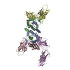
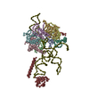
| Entry | Database: PDB / ID: 1knv | ||||||
|---|---|---|---|---|---|---|---|
| Title | Bse634I restriction endonuclease | ||||||
 Components Components | Bse634I restriction endonuclease | ||||||
 Keywords Keywords |  HYDROLASE / Restriction endonuclease / apo-enzyme HYDROLASE / Restriction endonuclease / apo-enzyme | ||||||
| Function / homology |  Function and homology information Function and homology information | ||||||
| Biological species |   Geobacillus stearothermophilus (bacteria) Geobacillus stearothermophilus (bacteria) | ||||||
| Method | X-RAY DIFFRACTION / SYNCHROTRON / MIR / Resolution: 2.17 Å | ||||||
 Authors Authors | Grazulis, S. / Deibert, M. / Rimseliene, R. / Skirgaila, R. / Sasnauskas, G. / Lagunavicius, A. / Repin, V. / Urbanke, C. / Huber, R. / Siksnys, V. | ||||||
 Citation Citation | Journal: Nucleic Acids Res. / Year: 2002 Title: Crystal structure of the Bse634I restriction endonuclease: comparison of two enzymes recognizing the same DNA sequence. Authors: Grazulis, S. / Deibert, M. / Rimseliene, R. / Skirgaila, R. / Sasnauskas, G. / Lagunavicius, A. / Repin, V. / Urbanke, C. / Huber, R. / Siksnys, V. | ||||||
| History |
|
- Structure visualization
| Structure viewer | Molecule: MolmilJmol/JSmol |
|---|
- Downloads & links
-Download
| PDBx/mmCIF format | 1knv.cif.gz | 132.5 KB | Display | PDBx/mmCIF format |
|---|---|---|---|---|
| PDB format | pdb1knv.ent.gz | 105.4 KB | Display | PDB format |
| PDBx/mmJSON format | 1knv.json.gz | Tree view | PDBx/mmJSON format | |
| Others |  Other downloads Other downloads |
-Validation report
| Arichive directory | https://data.pdbj.org/pub/pdb/validation_reports/kn/1knvftp://data.pdbj.org/pub/pdb/validation_reports/kn/1knv | HTTPS FTP |
|---|
-Related structure data
| Related structure data | |
|---|---|
| Similar structure data |


-Links
 PDBj
PDBj


- Assembly
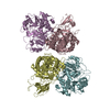
| Deposited unit | 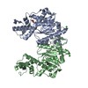
| ||||||||||||
|---|---|---|---|---|---|---|---|---|---|---|---|---|---|
| 1 | 
| ||||||||||||
| Unit cell |
| ||||||||||||
| Components on special symmetry positions |
| ||||||||||||
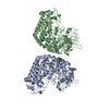
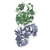
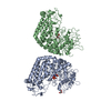
| Details | Biological assembly is a teramer (dimer of dimers); second dimer in the tetramer is generated by crystallographic two-fold axis -x+1, -y+1, z from the chains A and B |
-Components
| #1: Protein | Mass: 33848.875 Da / Num. of mol.: 2 Source method: isolated from a genetically manipulated source Source: (gene. exp.) Geobacillus stearothermophilus (bacteria)Plasmid: pUC18 / Production host: Escherichia coli (E. coli) / Strain (production host): ER2267References: UniProt: Q8RT53, type II site-specific deoxyribonuclease#2: Chemical | Acetate  Mass: 59.044 Da / Num. of mol.: 2 / Source method: obtained synthetically / Formula: C2H3O2 Mass: 59.044 Da / Num. of mol.: 2 / Source method: obtained synthetically / Formula: C2H3O2#3: Chemical | ChemComp-CL / Chloride  Mass: 35.453 Da / Num. of mol.: 5 / Source method: obtained synthetically / Formula: Cl Mass: 35.453 Da / Num. of mol.: 5 / Source method: obtained synthetically / Formula: Cl#4: Water | ChemComp-HOH / | Water Mass: 18.015 Da / Num. of mol.: 290 / Source method: isolated from a natural source / Formula: H2O Mass: 18.015 Da / Num. of mol.: 290 / Source method: isolated from a natural source / Formula: H2O |
|---|
-Experimental details
-Experiment
| Experiment | Method: X-RAY DIFFRACTION / Number of used crystals: 1 |
|---|
- Sample preparation
| Crystal | Density Matthews: 3.11 Å3/Da / Density % sol: 60.48 % | ||||||||||||||||||||||||||||||||||||||||||||||||||||||||
|---|---|---|---|---|---|---|---|---|---|---|---|---|---|---|---|---|---|---|---|---|---|---|---|---|---|---|---|---|---|---|---|---|---|---|---|---|---|---|---|---|---|---|---|---|---|---|---|---|---|---|---|---|---|---|---|---|---|
| Crystal grow | Temperature: 291 K / Method: vapor diffusion, sitting drop / pH: 5.5 Details: Na Acetate, PEG 8000, Calcium chloride, pH 5.5, VAPOR DIFFUSION, SITTING DROP, temperature 291K | ||||||||||||||||||||||||||||||||||||||||||||||||||||||||
| Crystal grow | *PLUS | ||||||||||||||||||||||||||||||||||||||||||||||||||||||||
| Components of the solutions | *PLUS
|
-Data collection
| Diffraction | Mean temperature: 90 K |
|---|---|
| Diffraction source | Source: SYNCHROTRON / Site: MPG/DESY, HAMBURG  / Beamline: BW6 / Wavelength: 1.09932 Å / Beamline: BW6 / Wavelength: 1.09932 Å |
| Detector | Type: MARRESEARCH / Detector: IMAGE PLATE / Date: Nov 2, 1997 / Details: monochromator, mirrors |
| Radiation | Monochromator: monochromator / Protocol: SINGLE WAVELENGTH / Monochromatic (M) / Laue (L): M / Scattering type: x-ray |
| Radiation wavelength | Wavelength: 1.09932 Å / Relative weight: 1 |
| Reflection | Resolution: 2.17→24.6 Å / Num. all: 43316 / Num. obs: 43275 / % possible obs: 95 % / Observed criterion σ(F): 0 / Observed criterion σ(I): 0 / Redundancy: 2.6 % / Biso Wilson estimate: 19.6 Å2 / Rmerge(I) obs: 0.055 |
| Reflection shell | Resolution: 2.17→2.23 Å / % possible all: 87.2 |
| Reflection | *PLUS Num. obs: 43316 |
- Processing
| Software |
| ||||||||||||||||||||||||||||||||||||
|---|---|---|---|---|---|---|---|---|---|---|---|---|---|---|---|---|---|---|---|---|---|---|---|---|---|---|---|---|---|---|---|---|---|---|---|---|---|
| Refinement | Method to determine structure: MIR / Resolution: 2.17→24.61 Å / Rfactor Rfree error: 0.004 / Data cutoff high absF: 2011306.16 / Data cutoff low absF: 0 / Isotropic thermal model: RESTRAINED / Cross valid method: THROUGHOUT / σ(F): 2 / σ(I): 0 / Stereochemistry target values: Engh & Huber
| ||||||||||||||||||||||||||||||||||||
| Solvent computation | Solvent model: FLAT MODEL / Bsol: 40.3493 Å2 / ksol: 0.3641 e/Å3 | ||||||||||||||||||||||||||||||||||||
| Displacement parameters | Biso mean: 32.7 Å2
| ||||||||||||||||||||||||||||||||||||
| Refine analyze |
| ||||||||||||||||||||||||||||||||||||
| Refinement step | Cycle: LAST / Resolution: 2.17→24.61 Å
| ||||||||||||||||||||||||||||||||||||
| Refine LS restraints |
| ||||||||||||||||||||||||||||||||||||
| LS refinement shell | Resolution: 2.17→2.3 Å / Rfactor Rfree error: 0.011 / Total num. of bins used: 6
| ||||||||||||||||||||||||||||||||||||
| Xplor file |
| ||||||||||||||||||||||||||||||||||||
| Software | *PLUS Name: CNS / Version: 1 / Classification: refinement | ||||||||||||||||||||||||||||||||||||
| Refinement | *PLUS Num. reflection obs: 43316 / σ(F): 2 / % reflection Rfree: 10 % / Rfactor Rfree: 0.252 | ||||||||||||||||||||||||||||||||||||
| Solvent computation | *PLUS | ||||||||||||||||||||||||||||||||||||
| Displacement parameters | *PLUS Biso mean: 32.7 Å2 | ||||||||||||||||||||||||||||||||||||
| Refine LS restraints | *PLUS
| ||||||||||||||||||||||||||||||||||||
| LS refinement shell | *PLUS Rfactor Rfree: 0.288 / % reflection Rfree: 10.5 % / Rfactor Rwork: 0.273 |
