[English] 日本語
Yorodumi
- PDB-4fix: Crystal Structure of GlfT2 -

+
Open data


ID or keywords:

Loading...

-
Basic information

Entry
Database: PDB / ID: 4fix
TitleCrystal Structure of GlfT2
ComponentsUDP-galactofuranosyl transferase GlfT2
Keywords
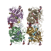
TRANSFERASE / Galactofuranosyltransferase / CAZY GT-2 family / Glycosyltransferase / Carbohydrate binding / Membrane
Function / homology
Function and homology information


galactofuranosylgalactofuranosylrhamnosyl-N-acetylglucosaminyl-diphospho-decaprenol beta-1,5/1,6-galactofuranosyltransferase / lipopolysaccharide-1,6-galactosyltransferase activity / lipopolysaccharide-1,5-galactosyltransferase activity / UDP-D-galactose metabolic process / cell wall macromolecule biosynthetic process / cell wall polysaccharide biosynthetic process / UDP-galactosyltransferase activity / mycolate cell wall layer assembly / capsule polysaccharide biosynthetic process / lipopolysaccharide biosynthetic process ...galactofuranosylgalactofuranosylrhamnosyl-N-acetylglucosaminyl-diphospho-decaprenol beta-1,5/1,6-galactofuranosyltransferase / lipopolysaccharide-1,6-galactosyltransferase activity / lipopolysaccharide-1,5-galactosyltransferase activity / UDP-D-galactose metabolic process / cell wall macromolecule biosynthetic process / cell wall polysaccharide biosynthetic process / UDP-galactosyltransferase activity / mycolate cell wall layer assembly / capsule polysaccharide biosynthetic process / lipopolysaccharide biosynthetic process / glycosyltransferase activity / cell wall organization / transferase activity / metal ion binding / plasma membrane / cytosol
Similarity search - Function
Spore Coat Polysaccharide Biosynthesis Protein SpsA; Chain A - #60 / Galactofuranosyltransferase GlfT2, N-terminal / Galactofuranosyltransferase-2, C-terminal / Galactofuranosyltransferase 2 N-terminal / Galactofuranosyltransferase-2, domain 3 / Glycosyltransferase like family 2 / Spore Coat Polysaccharide Biosynthesis Protein SpsA; Chain A / Nucleotide-diphospho-sugar transferases / Alpha-Beta Complex / Alpha Beta
Similarity search - Domain/homology
BETA-MERCAPTOETHANOL / : / Galactofuranosyltransferase GlfT2
Similarity search - Component
Biological speciesMycobacterium tuberculosis (bacteria)
MethodX-RAY DIFFRACTION / SYNCHROTRON / SAD / Resolution: 2.45 Å
AuthorsWheatley, R.W. / Zheng, R.B. / Lowary, T.L. / Ng, K.K.S.
CitationJournal: J.Biol.Chem. / Year: 2012
Title: Tetrameric Structure of the GlfT2 Galactofuranosyltransferase Reveals a Scaffold for the Assembly of Mycobacterial Arabinogalactan.
Authors: Wheatley, R.W. / Zheng, R.B. / Richards, M.R. / Lowary, T.L. / Ng, K.K.
History
DepositionJun 11, 2012Deposition site: RCSB / Processing site: RCSB
Revision 1.0Jun 20, 2012Provider: repository / Type: Initial release
Revision 1.1Aug 29, 2012Group: Database references
Revision 1.2Oct 17, 2012Group: Derived calculations
Revision 1.3Feb 28, 2024Group: Data collection / Database references / Derived calculations
Category: chem_comp_atom / chem_comp_bond ...chem_comp_atom / chem_comp_bond / database_2 / pdbx_struct_conn_angle / struct_conn / struct_ref_seq_dif / struct_site
Item: _database_2.pdbx_DOI / _database_2.pdbx_database_accession ..._database_2.pdbx_DOI / _database_2.pdbx_database_accession / _pdbx_struct_conn_angle.ptnr1_auth_comp_id / _pdbx_struct_conn_angle.ptnr1_auth_seq_id / _pdbx_struct_conn_angle.ptnr1_label_atom_id / _pdbx_struct_conn_angle.ptnr1_label_comp_id / _pdbx_struct_conn_angle.ptnr1_label_seq_id / _pdbx_struct_conn_angle.ptnr3_auth_comp_id / _pdbx_struct_conn_angle.ptnr3_auth_seq_id / _pdbx_struct_conn_angle.ptnr3_label_atom_id / _pdbx_struct_conn_angle.ptnr3_label_comp_id / _pdbx_struct_conn_angle.ptnr3_label_seq_id / _pdbx_struct_conn_angle.value / _struct_conn.pdbx_dist_value / _struct_conn.ptnr1_auth_asym_id / _struct_conn.ptnr1_auth_comp_id / _struct_conn.ptnr1_auth_seq_id / _struct_conn.ptnr1_label_asym_id / _struct_conn.ptnr1_label_atom_id / _struct_conn.ptnr1_label_comp_id / _struct_conn.ptnr1_label_seq_id / _struct_conn.ptnr2_auth_asym_id / _struct_conn.ptnr2_auth_seq_id / _struct_conn.ptnr2_label_asym_id / _struct_ref_seq_dif.details / _struct_site.pdbx_auth_asym_id / _struct_site.pdbx_auth_comp_id / _struct_site.pdbx_auth_seq_id

-
Structure visualization

Structure viewerMolecule:
MolmilJmol/JSmol

Downloads & links

-
Assembly

Deposited unit
A: UDP-galactofuranosyl transferase GlfT2
B: UDP-galactofuranosyl transferase GlfT2
hetero molecules


Theoretical massNumber of molelcules
Total (without water)148,0989
Polymers147,5422
Non-polymers5567
Water7,422412
1
A: UDP-galactofuranosyl transferase GlfT2
hetero molecules

A: UDP-galactofuranosyl transferase GlfT2
hetero molecules

A: UDP-galactofuranosyl transferase GlfT2
hetero molecules

A: UDP-galactofuranosyl transferase GlfT2
hetero molecules


Theoretical massNumber of molelcules
Total (without water)296,40820
Polymers295,0834
Non-polymers1,32516
Water724
TypeNameSymmetry operationNumber
identity operation1_555x,y,z1
crystal symmetry operation2_765-x+2,-y+1,z1
crystal symmetry operation3_645-y+3/2,x-1/2,z1
crystal symmetry operation4_565y+1/2,-x+3/2,z1
2
B: UDP-galactofuranosyl transferase GlfT2
hetero molecules

B: UDP-galactofuranosyl transferase GlfT2
hetero molecules

B: UDP-galactofuranosyl transferase GlfT2
hetero molecules

B: UDP-galactofuranosyl transferase GlfT2
hetero molecules


Theoretical massNumber of molelcules
Total (without water)295,98416
Polymers295,0834
Non-polymers90112
Water724
TypeNameSymmetry operationNumber
identity operation1_555x,y,z1
crystal symmetry operation2_655-x+1,-y,z1
crystal symmetry operation3_545-y+1/2,x-1/2,z1
crystal symmetry operation4_555y+1/2,-x+1/2,z1
Unit cell
Length a, b, c (Å)150.791, 150.791, 148.016
Angle α, β, γ (deg.)90.00, 90.00, 90.00
Int Tables number90
Space group name H-MP4212

-
Components

#1: Protein UDP-galactofuranosyl transferase GlfT2 / GalTr / Beta-D-(1-5)galactofuranosyltransferase / Beta-D-(1-6)galactofuranosyltransferase / UDP- ...GalTr / Beta-D-(1-5)galactofuranosyltransferase / Beta-D-(1-6)galactofuranosyltransferase / UDP-Galf:alpha-3-L-rhamnosyl-alpha-D-GlcNAc-pyrophosphate polyprenol / UDP-galactofuranosyl transferase


Mass: 73770.805 Da / Num. of mol.: 2
Source method: isolated from a genetically manipulated source
Source: (gene. exp.) Mycobacterium tuberculosis (bacteria) / Strain: H37Rv / Gene: glfT2, Rv3808c, RV38308c / Plasmid: pET-15B / Production host: Escherichia coli (E. coli) / Strain (production host): C41(DE3)
References: UniProt: O53585, Transferases; Glycosyltransferases; Hexosyltransferases
#2: Chemical
ChemComp-GOL / GLYCEROL / GLYCERIN / PROPANE-1,2,3-TRIOL / Glycerol


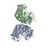
Mass: 92.094 Da / Num. of mol.: 4 / Source method: obtained synthetically / Formula: C3H8O3
#3: Chemical ChemComp-MN / MANGANESE (II) ION


Mass: 54.938 Da / Num. of mol.: 2 / Source method: obtained synthetically / Formula: Mn
#4: Chemical ChemComp-BME / BETA-MERCAPTOETHANOL / 2-Mercaptoethanol


Mass: 78.133 Da / Num. of mol.: 1 / Source method: obtained synthetically / Formula: C2H6OS
#5: Water ChemComp-HOH / water / Water


Mass: 18.015 Da / Num. of mol.: 412 / Source method: isolated from a natural source / Formula: H2O

-
Experimental details

-
Experiment

ExperimentMethod: X-RAY DIFFRACTION / Number of used crystals: 1

-
Sample preparation

CrystalDensity Matthews: 2.85 Å3/Da / Density % sol: 56.86 %
Crystal growTemperature: 298 K / Method: vapor diffusion, hanging drop / pH: 6.5
Details: 7% (w/v) PEG 4000, 100 mM sodium cacodylate pH 6.5, 100 mM sodium acetate, VAPOR DIFFUSION, HANGING DROP, temperature 298K

-
Data collection

DiffractionMean temperature: 100 K
Diffraction sourceSource: SYNCHROTRON / Site: SSRL / Beamline: BL9-2 / Wavelength: 1.0442 Å
DetectorType: MARMOSAIC 325 mm CCD / Detector: CCD / Date: Dec 18, 2008 / Details: mirrors
RadiationMonochromator: double crystal / Protocol: SINGLE WAVELENGTH / Monochromatic (M) / Laue (L): M / Scattering type: x-ray
Radiation wavelengthWavelength: 1.0442 Å / Relative weight: 1
ReflectionResolution: 2.45→40 Å / Num. all: 59863 / Num. obs: 59863 / % possible obs: 99.9 % / Observed criterion σ(F): -3 / Observed criterion σ(I): -3 / Redundancy: 11.9 % / Biso Wilson estimate: 56.2 Å2 / Rmerge(I) obs: 0.104 / Rsym value: 0.104 / Net I/σ(I): 27.2
Reflection shellResolution: 2.45→2.54 Å / Redundancy: 9.4 % / Rmerge(I) obs: 0.942 / Mean I/σ(I) obs: 2 / Num. unique all: 6192 / Rsym value: 0.942 / % possible all: 99.4

-
Processing

Software
NameVersionClassification
Blu-Icedata collection
PHENIXmodel building
REFMAC5.5.0109refinement
DENZOdata reduction
SCALEPACKdata scaling
PHENIXphasing
RefinementMethod to determine structure: SAD / Resolution: 2.45→40 Å / Cor.coef. Fo:Fc: 0.955 / Cor.coef. Fo:Fc free: 0.939 / SU B: 15.503 / SU ML: 0.162
Isotropic thermal model: isotropic atomic model plus TLS anisotropic model for domains
Cross valid method: THROUGHOUT / ESU R: 0.348 / ESU R Free: 0.231 / Stereochemistry target values: Engh & Huber
Details: TLSANL was used to determine the TLS-anisotropic components of temperature factors for all atoms as obtained by refining TLS parameters for individual domains
RfactorNum. reflection% reflectionSelection details
Rfree0.22228 3195 5.1 %RANDOM
Rwork0.18463 ---
all0.18657 59863 --
obs0.18657 59863 99.92 %-
Solvent computationIon probe radii: 0.8 Å / Shrinkage radii: 0.8 Å / VDW probe radii: 1.4 Å / Solvent model: BABINET MODEL WITH MASK
Displacement parametersBiso mean: 56.18 Å2
Baniso -1Baniso -2Baniso -3
1--1.45 Å20 Å20 Å2
2---1.45 Å20 Å2
3---2.91 Å2
Refinement stepCycle: LAST / Resolution: 2.45→40 Å
ProteinNucleic acidLigandSolventTotal
Num. atoms9966 0 30 412 10408
Refine LS restraints
Refine-IDTypeDev idealDev ideal targetNumber
X-RAY DIFFRACTIONr_bond_refined_d0.0080.02210259
X-RAY DIFFRACTIONr_angle_refined_deg1.0541.94713974
X-RAY DIFFRACTIONr_dihedral_angle_1_deg5.5251256
X-RAY DIFFRACTIONr_dihedral_angle_2_deg34.22922.75480
X-RAY DIFFRACTIONr_dihedral_angle_3_deg13.935151630
X-RAY DIFFRACTIONr_dihedral_angle_4_deg14.4741596
X-RAY DIFFRACTIONr_chiral_restr0.0730.21526
X-RAY DIFFRACTIONr_gen_planes_refined0.0040.0217930
X-RAY DIFFRACTIONr_mcbond_it1.28926300
X-RAY DIFFRACTIONr_mcangle_it2.2312.510176
X-RAY DIFFRACTIONr_scbond_it3.2453.53959
X-RAY DIFFRACTIONr_scangle_it4.9364.53798
LS refinement shellResolution: 2.451→2.514 Å / Total num. of bins used: 20
RfactorNum. reflection% reflection
Rfree0.296 229 -
Rwork0.243 4345 -
obs-4345 99.22 %
Refinement TLS params.

Method: refined / Refine-ID: X-RAY DIFFRACTION

IDL112)L122)L132)L222)L232)L332)S11 (Å °)S12 (Å °)S13 (Å °)S21 (Å °)S22 (Å °)S23 (Å °)S31 (Å °)S32 (Å °)S33 (Å °)T112)T122)T132)T222)T232)T332)Origin x (Å)Origin y (Å)Origin z (Å)
10.74430.03730.43610.87110.36971.0093-0.0306-0.1162-0.04170.04730.05350.11720.0302-0.1265-0.02290.0475-0.0189-0.00010.06830.03460.0525129.0662.99464.733
21.4639-0.24940.20171.4120.27370.95480.0773-0.083-0.45980.00040.05380.12620.18880.0018-0.13120.0762-0.0268-0.06490.02380.03740.1853132.63740.26753.361
35.3312-0.1934-1.79110.33150.01812.21630.23580.6595-0.6651-0.2604-0.20150.2730.2806-0.0397-0.03430.34420.1365-0.24020.2259-0.25330.3521140.26530.8331.001
43.00950.02921.08251.64321.81963.44810.20170.54170.0349-0.3912-0.10960.0726-0.28260.1464-0.09220.16830.1001-0.03310.178-0.06050.0778164.13943.34129.273
50.71250.318-0.20720.7567-0.48651.1419-0.0407-0.05420.00890.0136-0.0295-0.0305-0.04310.17170.07020.0453-0.01750.00890.07510.01020.037296.35113.36946.687
61.12950.1413-0.1111.3083-0.27111.3887-0.01210.06350.2067-0.03180.01310.0489-0.32470.0343-0.00110.1515-0.0459-0.01840.0410.04040.068991.90736.06535.44
74.84480.43921.16520.7142-0.57982.5484-0.11520.75860.3382-0.20160.0276-0.0395-0.3175-0.05270.08760.424-0.0267-0.02390.28380.18050.162983.8645.213.047
83.95591.3164-0.40312.7852-0.55254.0617-0.21450.995-0.1437-0.38180.28670.14380.1356-0.0979-0.07220.28990.0145-0.07620.39310.00870.06860.60331.46111.205
Refinement TLS group
IDRefine-IDRefine TLS-IDAuth asym-IDAuth seq-ID
1X-RAY DIFFRACTION1A1 - 157
2X-RAY DIFFRACTION2A158 - 405
3X-RAY DIFFRACTION3A406 - 492
4X-RAY DIFFRACTION3A580 - 629
5X-RAY DIFFRACTION4A493 - 579
6X-RAY DIFFRACTION5B1 - 157
7X-RAY DIFFRACTION6B158 - 405
8X-RAY DIFFRACTION7B406 - 492
9X-RAY DIFFRACTION7B580 - 629
10X-RAY DIFFRACTION8B493 - 579

+
About Yorodumi

-
News

-
Feb 9, 2022. New format data for meta-information of EMDB entries

New format data for meta-information of EMDB entries

  • Version 3 of the EMDB header file is now the official format.
  • The previous official version 1.9 will be removed from the archive.

Related info.:EMDB header

External links:wwPDB to switch to version 3 of the EMDB data model

-
Aug 12, 2020. Covid-19 info

Covid-19 info

URL: https://pdbj.org/emnavi/covid19.php

New page: Covid-19 featured information page in EM Navigator.

Related info.:Covid-19 info / Mar 5, 2020. Novel coronavirus structure data

+
Mar 5, 2020. Novel coronavirus structure data

Novel coronavirus structure data

Related info.:Yorodumi Speices / Aug 12, 2020. Covid-19 info

External links:COVID-19 featured content - PDBj / Molecule of the Month (242):Coronavirus Proteases

+
Jan 31, 2019. EMDB accession codes are about to change! (news from PDBe EMDB page)

EMDB accession codes are about to change! (news from PDBe EMDB page)

  • The allocation of 4 digits for EMDB accession codes will soon come to an end. Whilst these codes will remain in use, new EMDB accession codes will include an additional digit and will expand incrementally as the available range of codes is exhausted. The current 4-digit format prefixed with “EMD-” (i.e. EMD-XXXX) will advance to a 5-digit format (i.e. EMD-XXXXX), and so on. It is currently estimated that the 4-digit codes will be depleted around Spring 2019, at which point the 5-digit format will come into force.
  • The EM Navigator/Yorodumi systems omit the EMD- prefix.

Related info.:Q: What is EMD? / ID/Accession-code notation in Yorodumi/EM Navigator

External links:EMDB Accession Codes are Changing Soon! / Contact to PDBj

+
Jul 12, 2017. Major update of PDB

Major update of PDB

  • wwPDB released updated PDB data conforming to the new PDBx/mmCIF dictionary.
  • This is a major update changing the version number from 4 to 5, and with Remediation, in which all the entries are updated.
  • In this update, many items about electron microscopy experimental information are reorganized (e.g. em_software).
  • Now, EM Navigator and Yorodumi are based on the updated data.

External links:wwPDB Remediation / Enriched Model Files Conforming to OneDep Data Standards Now Available in the PDB FTP Archive

-
Yorodumi

Thousand views of thousand structures

  • Yorodumi is a browser for structure data from EMDB, PDB, SASBDB, etc.
  • This page is also the successor to EM Navigator detail page, and also detail information page/front-end page for Omokage search.
  • The word "yorodu" (or yorozu) is an old Japanese word meaning "ten thousand". "mi" (miru) is to see.

Related info.:EMDB / PDB / SASBDB / Comparison of 3 databanks / Yorodumi Search / Aug 31, 2016. New EM Navigator & Yorodumi / Yorodumi Papers / Jmol/JSmol / Function and homology information / Changes in new EM Navigator and Yorodumi

Read more