 Movie
Movie Controller

[English] 日本語
 Yorodumi
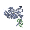
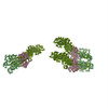
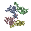
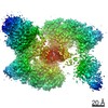
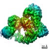
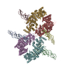
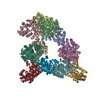
- PDB-1i84: CRYO-EM STRUCTURE OF THE HEAVY MEROMYOSIN SUBFRAGMENT OF CHICKEN ... -
+ Open data

- Basic information
| Entry | Database: PDB / ID: 1i84 | ||||||
|---|---|---|---|---|---|---|---|
| Title | CRYO-EM STRUCTURE OF THE HEAVY MEROMYOSIN SUBFRAGMENT OF CHICKEN GIZZARD SMOOTH MUSCLE MYOSIN WITH REGULATORY LIGHT CHAIN IN THE DEPHOSPHORYLATED STATE. ONLY C ALPHAS PROVIDED FOR REGULATORY LIGHT CHAIN. ONLY BACKBONE ATOMS PROVIDED FOR S2 FRAGMENT. | ||||||
 Components Components |
| ||||||
 Keywords Keywords | CONTRACTILE PROTEIN / muscle protein / smooth muscle / myosin subfragment 2 / heavy meromyosin / essential light chain / regulatory light chain / motor protein / coiled-coil | ||||||
| Function / homology |  Function and homology information Function and homology informationmyosin II filament / myofibril assembly / elastic fiber assembly / myosin light chain binding / skeletal muscle myosin thick filament assembly / myosin II binding / muscle myosin complex / actomyosin / myosin filament / actomyosin structure organization ...myosin II filament / myofibril assembly / elastic fiber assembly / myosin light chain binding / skeletal muscle myosin thick filament assembly / myosin II binding / muscle myosin complex / actomyosin / myosin filament / actomyosin structure organization / myosin II complex / myosin complex / cardiac muscle cell development / structural constituent of muscle / microfilament motor activity / myofibril / myosin heavy chain binding / smooth muscle contraction / muscle contraction / skeletal muscle tissue development / ADP binding / actin filament binding / actin binding / calmodulin binding / calcium ion binding / magnesium ion binding / ATP binding / cytoplasm / cytosol Similarity search - Function | ||||||
| Biological species |  | ||||||
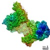
| Method | ELECTRON CRYSTALLOGRAPHY / electron crystallography / cryo EM / Resolution: 20 Å | ||||||
 Authors Authors | Wendt, T. / Taylor, D. / Trybus, K.M. / Taylor, K. | ||||||
 Citation Citation | Journal: Proc Natl Acad Sci U S A / Year: 2001 Title: Three-dimensional image reconstruction of dephosphorylated smooth muscle heavy meromyosin reveals asymmetry in the interaction between myosin heads and placement of subfragment 2. Authors: T Wendt / D Taylor / K M Trybus / K Taylor /  Abstract: Regulation of the actin-activated ATPase of smooth muscle myosin II is known to involve an interaction between the two heads that is controlled by phosphorylation of the regulatory light chain. ...Regulation of the actin-activated ATPase of smooth muscle myosin II is known to involve an interaction between the two heads that is controlled by phosphorylation of the regulatory light chain. However, the three-dimensional structure of this inactivated form has been unknown. We have used a lipid monolayer to obtain two-dimensional crystalline arrays of the unphosphorylated inactive form of smooth muscle heavy meromyosin suitable for structural studies by electron cryomicroscopy of unstained, frozen-hydrated specimens. The three-dimensional structure reveals an asymmetric interaction between the two myosin heads. The ATPase activity of one head is sterically "blocked" because part of its actin-binding interface is positioned onto the converter domain of the second head. ATPase activity of the second head, which can bind actin, appears to be inhibited through stabilization of converter domain movements needed to release phosphate and achieve strong actin binding. When the subfragment 2 domain of heavy meromyosin is oriented as it would be in an actomyosin filament lattice, the position of the heads is very different from that needed to bind actin, suggesting an additional contribution to ATPase inhibition in situ. #1: Journal: J.Cell Biol. / Year: 1999Title: Visualization of Head-head Interactions in the Inhibited State of Smooth Muscle Myosin Authors: Wendt, T. / Taylor, D. / Messier, T. / Trybus, K.M. / Taylor, K.A. #2: Journal: Cell(Cambridge,Mass.) / Year: 1998Title: Crystal Structure of a Vertebrate Smooth Muscle Myosin Motor Domain and its Complex with the Essential Light Chain: Visualization of the Pre-power Stroke State Authors: Dominguez, R. / Freyzon, Y. / Trybus, K.M. / Cohen, C. #3: Journal: Science / Year: 1993Title: Three-dimensional Structure of Myosin Subfragment-1: a Molecular Motor Authors: Rayment, I. / Rypniewsky, W.R. / Schmidt-Base, K. / Smith, R. / Tomchick, D.R. / Benning, M.M. / Winkelmann, D.A. / Wesenberg, G. / Holden, H.M. #4: Journal: J.Mol.Biol. / Year: 1996Title: The Structure of the Head-tail Junction of the Myosin Molecule Authors: Offer, G. / Knight, P. | ||||||
| History |
|
- Structure visualization
| Movie |
Movie viewer |
|---|---|
| Structure viewer | Molecule: MolmilJmol/JSmol |
- Downloads & links
-Download
| PDBx/mmCIF format | 1i84.cif.gz | 400.3 KB | Display | PDBx/mmCIF format |
|---|---|---|---|---|
| PDB format | pdb1i84.ent.gz | 313.6 KB | Display | PDB format |
| PDBx/mmJSON format | 1i84.json.gz | Tree view | PDBx/mmJSON format | |
| Others |  Other downloads Other downloads |
-Validation report
| Summary document | 1i84_validation.pdf.gz | 452.8 KB | Display | wwPDB validaton report |
|---|---|---|---|---|
| Full document | 1i84_full_validation.pdf.gz | 678.8 KB | Display | |
| Data in XML | 1i84_validation.xml.gz | 71.4 KB | Display | |
| Data in CIF | 1i84_validation.cif.gz | 104.2 KB | Display | |
| Arichive directory | https://data.pdbj.org/pub/pdb/validation_reports/i8/1i84ftp://data.pdbj.org/pub/pdb/validation_reports/i8/1i84 | HTTPS FTP |
-Related structure data
| Related structure data | |
|---|---|
| Similar structure data |

-Links
 PDBj
PDBj


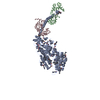
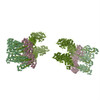
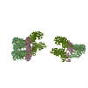
- Assembly
| Deposited unit | 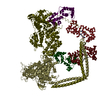
| ||||||||
|---|---|---|---|---|---|---|---|---|---|
| 1 |
| ||||||||
| Unit cell |
|
-Components
| #1: Antibody | Mass: 137072.938 Da / Num. of mol.: 2 / Fragment: MEROMYOSIN SUBFRAGMENT. S1 AND S2 FRAGMENTS. Source method: isolated from a genetically manipulated source Source: (gene. exp.)   Spodoptera frugiperda (fall armyworm) / Strain (production host): SF9 / References: UniProt: P10587, EC: 3.6.1.32 Spodoptera frugiperda (fall armyworm) / Strain (production host): SF9 / References: UniProt: P10587, EC: 3.6.1.32#2: Protein | Mass: 16873.025 Da / Num. of mol.: 2 / Fragment: S1 FRAGMENT Source method: isolated from a genetically manipulated source Source: (gene. exp.) Spodoptera frugiperda (fall armyworm) / Strain (production host): SF9 / References: GenBank: 212393, UniProt: P02607*PLUS#3: Protein | Mass: 18329.598 Da / Num. of mol.: 2 / Fragment: S1 FRAGMENT Source method: isolated from a genetically manipulated source Source: (gene. exp.) Spodoptera frugiperda (fall armyworm) / Strain (production host): SF9 / References: UniProt: P02609 |
|---|
-Experimental details
-Experiment
| Experiment | Method: ELECTRON CRYSTALLOGRAPHY / Number of used crystals: 101 |
|---|---|
| EM experiment | Aggregation state: 2D ARRAY / 3D reconstruction method: electron crystallography |
| Crystal symmetry | ∠γ: 91.5 ° / A: 133 Å / B: 304 Å / C: 90 Å / Space group name H-M: P2 |
- Sample preparation
| Component | Name: HEAVY MEROMYOSIN SUBFRAGMENT OF CHICKEN GIZZARD SMOOTH MUSCLE MYOSIN WITH REGULATORY LIGHT CHAIN IN THE DEPHOSPHORYLATED STATE Type: COMPLEX |
|---|---|
| EM crystal formation | Details: 2-D arrays were grown on a positively charged lipid monolayer system consisting of didodecyldimethylammonium and dilauryl phosphatidyl choline (Taylor, K. A. & Taylor, D. W. (1992) J. Struct. ...Details: 2-D arrays were grown on a positively charged lipid monolayer system consisting of didodecyldimethylammonium and dilauryl phosphatidyl choline (Taylor, K. A. & Taylor, D. W. (1992) J. Struct. Biol. 108, 140-147). Crystallization buffers consisted of 1 mM Mg2+, 20 mM phosphate, pH range 7-8, 1 mM ATP, 1 mM EGTA and PEG 6000 (7-10%) and NaCl (90-120 mM). Specimens were recovered on 200 mesh copper grids coated with a reticulated carbon film. The specimen was picked up from the grid bar side and blotted from that side. |
| Buffer solution | pH: 7.5 |
| Specimen | Embedding applied: NO / Shadowing applied: NO / Staining applied: NO / Vitrification applied: YES |
| Vitrification | Instrument: HOMEMADE PLUNGER / Cryogen name: ETHANE Details: Specimens were frozen in liquid ethane using a drop freezing apparatus. |
| Crystal grow | Temperature: 277 K / Method: lipid monolayer / pH: 7.5 Details: MgCl, sodium phosphate, ATP, EGTA, PEG 6000, pH 7.5, Lipid monolayer, temperature 277K |
| Crystal grow | *PLUS Method: other / Details: data none |
-Data collection
| Microscopy | Model: FEI/PHILIPS CM300FEG/T / Date: Jul 1, 1999 |
|---|---|
| Electron gun | Accelerating voltage: 300 kV / Illumination mode: FLOOD BEAM |
| Electron lens | Mode: BRIGHT FIELD / Nominal magnification: 24000 X |
| Specimen holder | Temperature: 93 K |
| Image recording | Electron dose: 5 e/Å2 / Film or detector model: KODAK SO-163 FILM |
| Image scans | Num. digital images: 101 / Scanner model: PERKIN ELMER |
| Radiation | Scattering type: electron |
| Radiation wavelength | Relative weight: 1 |
- Processing
| Image processing | Details: Micrographs were processed using the ICE processing software | ||||||||||||
|---|---|---|---|---|---|---|---|---|---|---|---|---|---|
| Crystal symmetry | ∠γ: 91.5 ° / A: 133 Å / B: 304 Å / C: 90 Å / Space group name H-M: P2 | ||||||||||||
| 3D reconstruction | Resolution: 20 Å / Resolution method: DIFFRACTION PATTERN/LAYERLINES / Symmetry type: 2D CRYSTAL | ||||||||||||
| Atomic model building | Protocol: OTHER / Space: REAL / Target criteria: VISUAL AGREEMENT Details: REFINEMENT PROTOCOL--MANUAL DETAILS--The atomic model consists of two S1 heads and a segment of S2 containing 91 residues. Each S1 consists of a heavy chain and two light chains, the ELC and ...Details: REFINEMENT PROTOCOL--MANUAL DETAILS--The atomic model consists of two S1 heads and a segment of S2 containing 91 residues. Each S1 consists of a heavy chain and two light chains, the ELC and the RLC. The atomic model of the S1 subfragment was constructed using two X-ray crystallography models. The heavy chain residues from ala2 to leu819 as well as the essential light chain were obtained from the chicken smooth muscle structure, PDB entry 1BR1, chains A and B. The remainder of S1 including heavy chain residues from glu820 to lys852 (smooth muscle numbering but skeletal myosin sequence) were obtained from residues glu811-lys843 of chicken skeletal muscle myosin, PDB entry 2MYS, since the structure of the entire smooth muscle S1 fragment is not available. Note that the residue numbers for the chain segment from 2MYS that were spliced onto 1BR1 have been renumbered to run consecutively from 819 but the residues themselves are the chicken skeletal muscle myosin heavy chain residues. The regulatory light chain coordinates were also obtained from 2MYS (skeletal) because a smooth muscle myosin model is not available. This regulatory light chain consisted only of C alphas. In fitting the model to the molecular envelope, the lever arm of the free head, i.e. ELC, RLC and heavy chain residues from ile796-lys852, were rotated as a rigid body about the Calpha of ile796. The amount of rotation was about 20 degrees. The blocked head was not modified. The alignment of 2MYS to 1BR1 was done using the conserved residues on the essential light chain. This alignment was carried out using O. The rms of the fit using 123 C alpha atoms of the conserved residues was 1.632 Angstroms. The C alpha distance between leu819 and glu811 (2MYS residue number) after the transformation was 4.23 Angstroms. This distance has not been altered in the model. The splice site at 819-820 is also slightly long and was left that way so the splice site would be easily visible. The other long bonds are at locations where the chain was bent to fit the structure. No minimization or refinement was done to fix them. The following residues have long peptide bond distances- chain S- LEU 819 and GLU 820 THR 854 and ARG 855 MET 860 and GLN 861 GLN 903 and ALA 904 chain V- LEU 819 and GLU 820 LYS 852 and VAL 853 MET 860 and GLN 861 GLN 903 and ALA 904 The 91 residues of the S2 fragment built into the model were calculated using a program written by Dr. Gerald Offer (reference 4). These coordinates consist only of backbone atoms. The S2 sequence begins at val853 which is the correct residue number for smooth muscle myosin. Peptide chain designations- Blocked myosin S1 head myosin heavy chain (with S2 segment) is chain V ELC is chain W RLC is chain Z Free myosin S1 head myosin heavy chain (with S2 fragment) is chain S ELC is chain T RLC is chain U The terms blocked and free refer to the conformations of the two S1 heads. SEQUENCE GAPS IN THE MOLECULAR MODEL- Myosin heavy chain- 205-210, 452-457, 635-655, 944-1185. ELC- 1-2. RLC (skeletal)- 1-18, 127, 142-147, 164-166. | ||||||||||||
| Refinement | Highest resolution: 20 Å | ||||||||||||
| Refinement step | Cycle: LAST / Highest resolution: 20 Å
|
