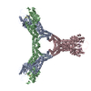
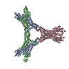
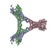
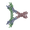
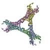
Journal: Nat Commun / Year: 2019 Title: Structure of Csx1-cOA complex reveals the basis of RNA decay in Type III-B CRISPR-Cas. Authors: Rafael Molina / Stefano Stella / Mingxia Feng / Nicholas Sofos / Vykintas Jauniskis / Irina Pozdnyakova / Blanca López-Méndez / Qunxin She / Guillermo Montoya / Abstract: Type III CRISPR-Cas multisubunit complexes cleave ssRNA and ssDNA. These activities promote the generation of cyclic oligoadenylate (cOA), which activates associated CRISPR-Cas RNases from the ...Type III CRISPR-Cas multisubunit complexes cleave ssRNA and ssDNA. These activities promote the generation of cyclic oligoadenylate (cOA), which activates associated CRISPR-Cas RNases from the Csm/Csx families, triggering a massive RNA decay to provide immunity from genetic invaders. Here we present the structure of Sulfolobus islandicus (Sis) Csx1-cOA complex revealing the allosteric activation of its RNase activity. SisCsx1 is a hexamer built by a trimer of dimers. Each dimer forms a cOA binding site and a ssRNA catalytic pocket. cOA undergoes a conformational change upon binding in the second messenger binding site activating ssRNA degradation in the catalytic pockets. Activation is transmitted in an allosteric manner through an intermediate HTH domain, which joins the cOA and catalytic sites. The RNase functions in a sequential cooperative fashion, hydrolyzing phosphodiester bonds in 5'-C-C-3'. The degradation of cOA by Ring nucleases deactivates SisCsx1, suggesting that this enzyme could be employed in biotechnological applications.
A: CRISPR-associated (Cas) DxTHG family B: CRISPR-associated (Cas) DxTHG family C: CRISPR-associated (Cas) DxTHG family D: CRISPR-associated (Cas) DxTHG family E: CRISPR-associated (Cas) DxTHG family F: CRISPR-associated (Cas) DxTHG family
In the structure databanks used in Yorodumi, some data are registered as the other names, "COVID-19 virus" and "2019-nCoV". Here are the details of the virus and the list of structure data.
Jan 31, 2019. EMDB accession codes are about to change! (news from PDBe EMDB page)
EMDB accession codes are about to change! (news from PDBe EMDB page)
The allocation of 4 digits for EMDB accession codes will soon come to an end. Whilst these codes will remain in use, new EMDB accession codes will include an additional digit and will expand incrementally as the available range of codes is exhausted. The current 4-digit format prefixed with “EMD-” (i.e. EMD-XXXX) will advance to a 5-digit format (i.e. EMD-XXXXX), and so on. It is currently estimated that the 4-digit codes will be depleted around Spring 2019, at which point the 5-digit format will come into force.
The EM Navigator/Yorodumi systems omit the EMD- prefix.
Related info.:Q: What is EMD? / ID/Accession-code notation in Yorodumi/EM Navigator
Yorodumi is a browser for structure data from EMDB, PDB, SASBDB, etc.
This page is also the successor to EM Navigator detail page, and also detail information page/front-end page for Omokage search.
The word "yorodu" (or yorozu) is an old Japanese word meaning "ten thousand". "mi" (miru) is to see.
Related info.:EMDB / PDB / SASBDB / Comparison of 3 databanks / Yorodumi Search / Aug 31, 2016. New EM Navigator & Yorodumi / Yorodumi Papers / Jmol/JSmol / Function and homology information / Changes in new EM Navigator and Yorodumi
 Movie
Movie Controller

 Yorodumi
 Open data
 Basic information
 Components
 Keywords
 RNA BINDING PROTEIN / CRISPR ASSOCIATED PROTEIN CARF HEPN RNAse
 Function and homology information

 Authors
 Denmark, 1items
  Citation
 Structure visualization
 Downloads & links
 Other downloads


 PDBj
PDBj Assembly
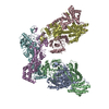
 Sample preparation
 / Beamline: X06SA / Wavelength: 1 Å
 Processing