 Movie
Movie Controller

[English] 日本語
 Yorodumi
- PDB-1gp4: Anthocyanidin synthase from Arabidopsis thaliana (selenomethionin... -
+ Open data

- Basic information
| Entry | Database: PDB / ID: 1gp4 | ||||||
|---|---|---|---|---|---|---|---|
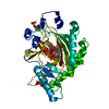
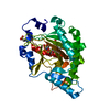
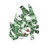
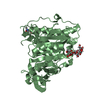
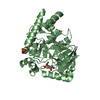
| Title | Anthocyanidin synthase from Arabidopsis thaliana (selenomethionine substituted) | ||||||
 Components Components | ANTHOCYANIDIN SYNTHASE Leucocyanidin oxygenase Leucocyanidin oxygenase | ||||||
 Keywords Keywords | OXIDOREDUCTASE / OXYGENASE / 2-OXOGLUTARATE DEPENDENT DIOXYGENASE / FLAVONOID BIOSYNTHESIS | ||||||
| Function / homology |  Function and homology informationanthocyanidin synthase / proanthocyanidin biosynthetic process / leucocyanidin oxygenase activity / anthocyanin-containing compound biosynthetic process / response to jasmonic acid / vacuole organization / L-ascorbic acid binding / response to wounding / metal ion binding Function and homology informationanthocyanidin synthase / proanthocyanidin biosynthetic process / leucocyanidin oxygenase activity / anthocyanin-containing compound biosynthetic process / response to jasmonic acid / vacuole organization / L-ascorbic acid binding / response to wounding / metal ion bindingSimilarity search - Function | ||||||
| Biological species |  ARABIDOPSIS THALIANA (thale cress) ARABIDOPSIS THALIANA (thale cress) | ||||||
| Method | X-RAY DIFFRACTION / SYNCHROTRON / MAD / Resolution: 2.1 Å | ||||||
 Authors Authors | Wilmouth, R.C. / Turnbull, J.J. / Welford, R.W.D. / Clifton, I.J. / Prescott, A.G. / Schofield, C.J. | ||||||
 Citation Citation | Journal: Structure / Year: 2002 Title: Structure and Mechanism of Anthocyanidin Synthase from Arabidopsis Thaliana. Authors: Wilmouth, R.C. / Turnbull, J.J. / Welford, R.W.D. / Clifton, I.J. / Prescott, A.G. / Schofield, C.J. #1: Journal: Chem.Commun. / Year: 2000Title: Are Anthocyanidins the Immediate Products of Anthocyanidin Synthase? Authors: Turnbull, J.J. / Sobey, W.J. / Aplin, R.T. / Hassan, A. / Firmin, J.L. / Schofield, C.J. / Prescott, A.G. #2: Journal: Acta Crystallogr.,Sect.D / Year: 2001 Title: Purification, Crystallization and Preliminary X-Ray Diffraction of Anthocyanidin Synthase from Arabidopsis Thaliana Authors: Turnbull, J.J. / Prescott, A.G. / Schofield, C.J. / Wilmouth, R.C. #3: Journal: Plant J. / Year: 1999 Title: Direct Evidence for Anthocyanidin Synthase as a 2-Oxoglutarate-Dependent Oxygenase: Molecular Cloning and Functional Expression of Cdna from a Red Forma of Perilla Frutescens Authors: Saito, K. / Kobayashi, M. / Gong, Z. / Tanaka, Y. / Yamazaki, M. | ||||||
| History |
|
- Structure visualization
| Structure viewer | Molecule: MolmilJmol/JSmol |
|---|
- Downloads & links
-Download
| PDBx/mmCIF format | 1gp4.cif.gz | 84.3 KB | Display | PDBx/mmCIF format |
|---|---|---|---|---|
| PDB format | pdb1gp4.ent.gz | 67.3 KB | Display | PDB format |
| PDBx/mmJSON format | 1gp4.json.gz | Tree view | PDBx/mmJSON format | |
| Others |  Other downloads Other downloads |
-Validation report
| Arichive directory | https://data.pdbj.org/pub/pdb/validation_reports/gp/1gp4ftp://data.pdbj.org/pub/pdb/validation_reports/gp/1gp4 | HTTPS FTP |
|---|
-Related structure data


-Links
 PDBj
PDBj

- Assembly
| Deposited unit | 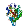
| ||||||||
|---|---|---|---|---|---|---|---|---|---|
| 1 |
| ||||||||
| Unit cell |
|
-Components
| #1: Protein | Leucocyanidin oxygenase / LEUCOANTHOCYANIDIN DIOXYGENASE Mass: 40779.617 Da / Num. of mol.: 1 Source method: isolated from a genetically manipulated source Source: (gene. exp.) ARABIDOPSIS THALIANA (thale cress) / Plasmid: PET-24A / Production host:  ESCHERICHIA COLI (E. coli) / Strain (production host): BL21(DE3) / References: UniProt: Q96323, EC: 1.14.11.19 ESCHERICHIA COLI (E. coli) / Strain (production host): BL21(DE3) / References: UniProt: Q96323, EC: 1.14.11.19 |
|---|---|
| #2: Chemical | ChemComp-AKG / Α-Ketoglutaric acid  Mass: 146.098 Da / Num. of mol.: 1 / Source method: obtained synthetically / Formula: C5H6O5 Mass: 146.098 Da / Num. of mol.: 1 / Source method: obtained synthetically / Formula: C5H6O5 |
| #3: Chemical | ChemComp-MES / MES (buffer)  Mass: 195.237 Da / Num. of mol.: 1 / Source method: obtained synthetically / Formula: C6H13NO4S / Comment: pH buffer*YM Mass: 195.237 Da / Num. of mol.: 1 / Source method: obtained synthetically / Formula: C6H13NO4S / Comment: pH buffer*YM |
| #4: Water | ChemComp-HOH / Water Mass: 18.015 Da / Num. of mol.: 288 / Source method: isolated from a natural source / Formula: H2O Mass: 18.015 Da / Num. of mol.: 288 / Source method: isolated from a natural source / Formula: H2O |
-Experimental details
-Experiment
| Experiment | Method: X-RAY DIFFRACTION / Number of used crystals: 1 |
|---|
- Sample preparation
| Crystal | Density Matthews: 2.41 Å3/Da / Density % sol: 48.6 % | ||||||||||||||||||||||||||||||||||||||||||||||||||||||||
|---|---|---|---|---|---|---|---|---|---|---|---|---|---|---|---|---|---|---|---|---|---|---|---|---|---|---|---|---|---|---|---|---|---|---|---|---|---|---|---|---|---|---|---|---|---|---|---|---|---|---|---|---|---|---|---|---|---|
| Crystal grow | pH: 6.5 Details: 30% (W/V) PEG 2000 MONOMETHYLETHER, 50 MM MES, 100 MM SODIUM CITRATE, 200 MM AMMONIUM ACETATE, 5 MM IRON(II) SULPHATE, 10 MM POTASSIUM ALPHA-KETOGLUTARATE, 10 MM SODIUM ASCORBATE, PH 6.5, ...Details: 30% (W/V) PEG 2000 MONOMETHYLETHER, 50 MM MES, 100 MM SODIUM CITRATE, 200 MM AMMONIUM ACETATE, 5 MM IRON(II) SULPHATE, 10 MM POTASSIUM ALPHA-KETOGLUTARATE, 10 MM SODIUM ASCORBATE, PH 6.5, ANAEROBIC (AR ATMOSPHERE, < 0.5 PPM OXYGEN) | ||||||||||||||||||||||||||||||||||||||||||||||||||||||||
| Crystal grow | *PLUS Method: vapor diffusion, hanging dropDetails: Turnbull, J.J., (2001) Acta Crystallogr.,Sect.D, D57, 425. | ||||||||||||||||||||||||||||||||||||||||||||||||||||||||
| Components of the solutions | *PLUS
|
-Data collection
| Diffraction | Mean temperature: 100 K | ||||||||||||
|---|---|---|---|---|---|---|---|---|---|---|---|---|---|
| Diffraction source | Source: SYNCHROTRON / Site: ESRF  / Beamline: BM14 / Wavelength: 0.91841,0.97855,0.97885 / Beamline: BM14 / Wavelength: 0.91841,0.97855,0.97885 | ||||||||||||
| Detector | Type: MARRESEARCH / Detector: CCD / Date: Sep 15, 2000 / Details: MIRRORS | ||||||||||||
| Radiation | Monochromator: SI / Protocol: MAD / Monochromatic (M) / Laue (L): M / Scattering type: x-ray | ||||||||||||
| Radiation wavelength |
| ||||||||||||
| Reflection | Resolution: 2.1→36.5 Å / Num. obs: 23462 / % possible obs: 99.9 % / Redundancy: 5.4 % / Biso Wilson estimate: 27.3 Å2 / Rmerge(I) obs: 0.063 / Net I/σ(I): 9.8 | ||||||||||||
| Reflection shell | Resolution: 2.1→2.21 Å / Rmerge(I) obs: 0.251 / Mean I/σ(I) obs: 2.8 / % possible all: 100 | ||||||||||||
| Reflection | *PLUS Num. measured all: 348658 | ||||||||||||
| Reflection shell | *PLUS Highest resolution: 2.1 Å / % possible obs: 100 % |
- Processing
| Software |
| ||||||||||||||||||||||||||||||||||||||||||||||||||||||||||||||||||||||||||||||||
|---|---|---|---|---|---|---|---|---|---|---|---|---|---|---|---|---|---|---|---|---|---|---|---|---|---|---|---|---|---|---|---|---|---|---|---|---|---|---|---|---|---|---|---|---|---|---|---|---|---|---|---|---|---|---|---|---|---|---|---|---|---|---|---|---|---|---|---|---|---|---|---|---|---|---|---|---|---|---|---|---|---|
| Refinement | Method to determine structure: MAD / Resolution: 2.1→20 Å / Rfactor Rfree error: 0.008 / Data cutoff high absF: 1571181.72 / Isotropic thermal model: RESTRAINED / Cross valid method: THROUGHOUT / σ(F): 0
| ||||||||||||||||||||||||||||||||||||||||||||||||||||||||||||||||||||||||||||||||
| Solvent computation | Solvent model: FLAT MODEL / Bsol: 46.5 Å2 / ksol: 0.336 e/Å3 | ||||||||||||||||||||||||||||||||||||||||||||||||||||||||||||||||||||||||||||||||
| Displacement parameters | Biso mean: 26.9 Å2
| ||||||||||||||||||||||||||||||||||||||||||||||||||||||||||||||||||||||||||||||||
| Refine analyze |
| ||||||||||||||||||||||||||||||||||||||||||||||||||||||||||||||||||||||||||||||||
| Refinement step | Cycle: LAST / Resolution: 2.1→20 Å
| ||||||||||||||||||||||||||||||||||||||||||||||||||||||||||||||||||||||||||||||||
| Refine LS restraints |
| ||||||||||||||||||||||||||||||||||||||||||||||||||||||||||||||||||||||||||||||||
| LS refinement shell | Resolution: 2.1→2.23 Å / Rfactor Rfree error: 0.02 / Total num. of bins used: 6
| ||||||||||||||||||||||||||||||||||||||||||||||||||||||||||||||||||||||||||||||||
| Xplor file |
| ||||||||||||||||||||||||||||||||||||||||||||||||||||||||||||||||||||||||||||||||
| Software | *PLUS Name: CNS / Version: 1.1 / Classification: refinement | ||||||||||||||||||||||||||||||||||||||||||||||||||||||||||||||||||||||||||||||||
| Refine LS restraints | *PLUS
| ||||||||||||||||||||||||||||||||||||||||||||||||||||||||||||||||||||||||||||||||
| LS refinement shell | *PLUS Rfactor obs: 0.192 |
