 Movie
Movie Controller

[English] 日本語
 Yorodumi
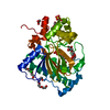
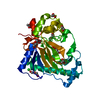
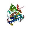
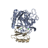
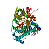
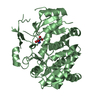
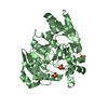
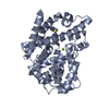
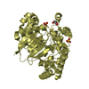
- PDB-5y9i: Crystal structure of the Kdo hydroxylase KdoO, a non-heme Fe(II) ... -
+ Open data

- Basic information
| Entry | Database: PDB / ID: 5y9i | ||||||
|---|---|---|---|---|---|---|---|
| Title | Crystal structure of the Kdo hydroxylase KdoO, a non-heme Fe(II) alphaketoglutarate dependent dioxygenase in complex with Co(II) | ||||||
 Components Components | Uncharacterized protein KdoO | ||||||
 Keywords Keywords |  BIOSYNTHETIC PROTEIN / Fe(II)/O2/alphaketoglutarate dependent dioxygenase / KDO2-lipid A dioxygenase / deoxysugar dioxygenase / LPS biosynthesis BIOSYNTHETIC PROTEIN / Fe(II)/O2/alphaketoglutarate dependent dioxygenase / KDO2-lipid A dioxygenase / deoxysugar dioxygenase / LPS biosynthesis | ||||||
| Function / homology | Kdo hydroxylase / 3-deoxy-D-manno-oct-2-ulosonic acid (Kdo) hydroxylase / ACETATE ION / : / 3-deoxy-D-manno-oct-2-ulosonic acid (Kdo) hydroxylase Function and homology information Function and homology information | ||||||
| Biological species |  Methylacidiphilum infernorum (bacteria) Methylacidiphilum infernorum (bacteria) | ||||||
| Method | X-RAY DIFFRACTION / SYNCHROTRON / SAD / Resolution: 1.901 Å | ||||||
 Authors Authors | Chung, H.S. / Pemble, C.W. / Joo, S.H. | ||||||
 Citation Citation | Journal: To Be Published Title: Biochemical and structural insights of Fe(II)/alpha-ketoglutarate/O2-dependent dioxygenase, KdoO from Methylacidiphilum infernorum V4 Authors: Chung, H.S. / Pemble IV, C.W. / Joo, S.H. | ||||||
| History |
|
- Structure visualization
| Structure viewer | Molecule: MolmilJmol/JSmol |
|---|
- Downloads & links
-Download
| PDBx/mmCIF format | 5y9i.cif.gz | 85.8 KB | Display | PDBx/mmCIF format |
|---|---|---|---|---|
| PDB format | pdb5y9i.ent.gz | 61.4 KB | Display | PDB format |
| PDBx/mmJSON format | 5y9i.json.gz | Tree view | PDBx/mmJSON format | |
| Others |  Other downloads Other downloads |
-Validation report
| Arichive directory | https://data.pdbj.org/pub/pdb/validation_reports/y9/5y9iftp://data.pdbj.org/pub/pdb/validation_reports/y9/5y9i | HTTPS FTP |
|---|
-Related structure data


-Links
 PDBj
PDBj
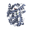
- Assembly
| Deposited unit | 
| ||||||||
|---|---|---|---|---|---|---|---|---|---|
| 1 |
| ||||||||
| Unit cell |
|
-Components
-Protein / Sugars , 2 types, 2 molecules A
| #1: Protein | Mass: 36239.316 Da / Num. of mol.: 1 Source method: isolated from a genetically manipulated source Source: (gene. exp.) Methylacidiphilum infernorum (isolate V4) (bacteria)Strain: isolate V4 / Gene: Minf_1012 / Production host: Escherichia coli (E. coli) / References: UniProt: B3DUR4 |
|---|---|
| #4: Sugar | ChemComp-BOG / Octyl glucoside Type: D-saccharide / Mass: 292.369 Da / Num. of mol.: 1 Type: D-saccharide / Mass: 292.369 Da / Num. of mol.: 1Source method: isolated from a genetically manipulated source Formula: C14H28O6 / Comment: detergent*YM |
-Non-polymers , 5 types, 398 molecules 


| #2: Chemical | ChemComp-SO4 / Sulfate Mass: 96.063 Da / Num. of mol.: 1 / Source method: obtained synthetically / Formula: SO4 Mass: 96.063 Da / Num. of mol.: 1 / Source method: obtained synthetically / Formula: SO4 | ||||
|---|---|---|---|---|---|
| #3: Chemical | ChemComp-CO /  Mass: 58.933 Da / Num. of mol.: 1 / Source method: obtained synthetically / Formula: Co Mass: 58.933 Da / Num. of mol.: 1 / Source method: obtained synthetically / Formula: Co | ||||
| #5: Chemical | Chloride Mass: 35.453 Da / Num. of mol.: 3 / Source method: obtained synthetically / Formula: Cl Mass: 35.453 Da / Num. of mol.: 3 / Source method: obtained synthetically / Formula: Cl#6: Chemical | Acetate Mass: 59.044 Da / Num. of mol.: 2 / Source method: obtained synthetically / Formula: C2H3O2 Mass: 59.044 Da / Num. of mol.: 2 / Source method: obtained synthetically / Formula: C2H3O2#7: Water | ChemComp-HOH / | WaterMass: 18.015 Da / Num. of mol.: 391 / Source method: isolated from a natural source / Formula: H2O |
-Experimental details
-Experiment
| Experiment | Method: X-RAY DIFFRACTION / Number of used crystals: 1 |
|---|
- Sample preparation
| Crystal | Density Matthews: 2.22 Å3/Da / Density % sol: 44.68 % |
|---|---|
| Crystal grow | Temperature: 288 K / Method: vapor diffusion, sitting drop Details: 0.1M Sodium acetate (pH=4.6), 200mM Ammonium sulfate, 25% v/v PEG 4000 |
-Data collection
| Diffraction | Mean temperature: 100 K |
|---|---|
| Diffraction source | Source: SYNCHROTRON / Site: APS  / Beamline: 22-BM / Wavelength: 1 Å / Beamline: 22-BM / Wavelength: 1 Å |
| Detector | Type: MARMOSAIC 225 mm CCD / Detector: CCD / Date: Jun 23, 2011 |
| Radiation | Monochromator: Si(111) / Protocol: SINGLE WAVELENGTH / Monochromatic (M) / Laue (L): M / Scattering type: x-ray |
| Radiation wavelength | Wavelength: 1 Å / Relative weight: 1 |
| Reflection | Resolution: 1.9→50 Å / Num. obs: 51868 / % possible obs: 98.3 % / Redundancy: 9.4 % / Net I/σ(I): 19.2 |
- Processing
| Software |
| ||||||||||||||||||||||||||||||||||||||||||||||||||||||||||||||||||||||||||||||||||||||||||||||||||||||||||||||||||||||||||||||
|---|---|---|---|---|---|---|---|---|---|---|---|---|---|---|---|---|---|---|---|---|---|---|---|---|---|---|---|---|---|---|---|---|---|---|---|---|---|---|---|---|---|---|---|---|---|---|---|---|---|---|---|---|---|---|---|---|---|---|---|---|---|---|---|---|---|---|---|---|---|---|---|---|---|---|---|---|---|---|---|---|---|---|---|---|---|---|---|---|---|---|---|---|---|---|---|---|---|---|---|---|---|---|---|---|---|---|---|---|---|---|---|---|---|---|---|---|---|---|---|---|---|---|---|---|---|---|---|
| Refinement | Method to determine structure: SAD / Resolution: 1.901→34.875 Å / SU ML: 0.16 / Cross valid method: FREE R-VALUE / σ(F): 1.35 / Phase error: 17.61 Details: THE STRUCTURE FACTOR FILE CONTAINS FRIEDEL PAIRS IN I_PLUS/MINUS COLUMNS
| ||||||||||||||||||||||||||||||||||||||||||||||||||||||||||||||||||||||||||||||||||||||||||||||||||||||||||||||||||||||||||||||
| Solvent computation | Shrinkage radii: 0.9 Å / VDW probe radii: 1.11 Å | ||||||||||||||||||||||||||||||||||||||||||||||||||||||||||||||||||||||||||||||||||||||||||||||||||||||||||||||||||||||||||||||
| Refinement step | Cycle: LAST / Resolution: 1.901→34.875 Å
| ||||||||||||||||||||||||||||||||||||||||||||||||||||||||||||||||||||||||||||||||||||||||||||||||||||||||||||||||||||||||||||||
| Refine LS restraints |
| ||||||||||||||||||||||||||||||||||||||||||||||||||||||||||||||||||||||||||||||||||||||||||||||||||||||||||||||||||||||||||||||
| LS refinement shell |
|
