 Movie
Movie Controller

[English] 日本語
 Yorodumi
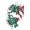
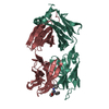
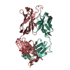
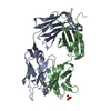
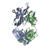
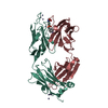
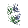
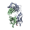
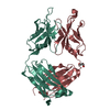
- PDB-1etz: THE THREE-DIMENSIONAL STRUCTURE OF AN ANTI-SWEETENER FAB, NC10.14... -
+ Open data

- Basic information
| Entry | Database: PDB / ID: 1etz | ||||||
|---|---|---|---|---|---|---|---|
| Title | THE THREE-DIMENSIONAL STRUCTURE OF AN ANTI-SWEETENER FAB, NC10.14, SHOWS THE EXTENT OF STRUCTURAL DIVERSITY IN ANTIGEN RECOGNITION BY IMMUNOGLOBULINS | ||||||
 Components Components |
| ||||||
 Keywords Keywords |  IMMUNE SYSTEM / Anti-sweetener Fab / Antigen-antibody / Complex / Receptor Mimicry / Antigen Recognition IMMUNE SYSTEM / Anti-sweetener Fab / Antigen-antibody / Complex / Receptor Mimicry / Antigen Recognition | ||||||
| Function / homology |  Function and homology information Function and homology informationInitial triggering of complement / Classical antibody-mediated complement activation / FCGR activation / Role of phospholipids in phagocytosis / Regulation of Complement cascade / Regulation of actin dynamics for phagocytic cup formation / humoral immune response mediated by circulating immunoglobulin / phagocytosis, recognition / positive regulation of type IIa hypersensitivity / positive regulation of type I hypersensitivity ...Initial triggering of complement / Classical antibody-mediated complement activation / FCGR activation / Role of phospholipids in phagocytosis / Regulation of Complement cascade / Regulation of actin dynamics for phagocytic cup formation / humoral immune response mediated by circulating immunoglobulin / phagocytosis, recognition / positive regulation of type IIa hypersensitivity / positive regulation of type I hypersensitivity / phagocytosis, engulfment / immunoglobulin mediated immune response / immunoglobulin complex, circulating / immunoglobulin receptor binding / positive regulation of phagocytosis / complement activation, classical pathway / antigen binding / positive regulation of immune response / antibacterial humoral response / blood microparticle / extracellular space / extracellular exosome / extracellular region / plasma membraneSimilarity search - Function | ||||||
| Biological species |  Mus musculus (house mouse) Mus musculus (house mouse) | ||||||
| Method | X-RAY DIFFRACTION / Resolution: 2.6 Å | ||||||
 Authors Authors | Guddat, L.W. / Shan, L. / Broomell, C. / Ramsland, P.A. / Fan, Z. / Anchin, J.M. / Linthicum, D.S. / Edmundson, A.B. | ||||||
 Citation Citation | Journal: J.Mol.Biol. / Year: 2000 Title: The three-dimensional structure of a complex of a murine Fab (NC10. 14) with a potent sweetener (NC174): an illustration of structural diversity in antigen recognition by immunoglobulins. Authors: Guddat, L.W. / Shan, L. / Broomell, C. / Ramsland, P.A. / Fan, Z. / Anchin, J.M. / Linthicum, D.S. / Edmundson, A.B. #1: Journal: J.Mol.Biol. / Year: 1994Title: Local and Transmitted Conformational Changes on Complexation of an Anti-sweetener Fab Authors: Guddat, L.W. / Shan, L. / Anchin, J.M. / Linthicum, D.S. / Edmundson, A.B. | ||||||
| History |
|
- Structure visualization
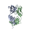
| Structure viewer | Molecule: MolmilJmol/JSmol |
|---|
- Downloads & links
-Download
| PDBx/mmCIF format | 1etz.cif.gz | 173.2 KB | Display | PDBx/mmCIF format |
|---|---|---|---|---|
| PDB format | pdb1etz.ent.gz | 144.2 KB | Display | PDB format |
| PDBx/mmJSON format | 1etz.json.gz | Tree view | PDBx/mmJSON format | |
| Others |  Other downloads Other downloads |
-Validation report
| Arichive directory | https://data.pdbj.org/pub/pdb/validation_reports/et/1etzftp://data.pdbj.org/pub/pdb/validation_reports/et/1etz | HTTPS FTP |
|---|
-Related structure data
| Related structure data | |
|---|---|
| Similar structure data |


-Links
 PDBj
PDBj


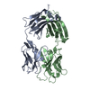
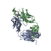
- Assembly
| Deposited unit | 
| ||||||||
|---|---|---|---|---|---|---|---|---|---|
| 1 | 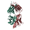
| ||||||||
| 2 | 
| ||||||||
| Unit cell |
| ||||||||
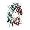
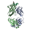
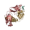
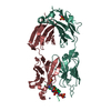
| Details | A Fab is formed by Light Chain and Heavy Chain. There are two Fab with non-crystallographic symmetry in a unit cell. |
-Components
| #1: Antibody | Mass: 23165.719 Da / Num. of mol.: 2 / Source method: isolated from a natural source / Source: (natural) Mus musculus (house mouse) / Tissue: ASCITES / References: UniProt: G0YP42*PLUS#2: Antibody | Mass: 24468.146 Da / Num. of mol.: 2 / Source method: isolated from a natural source / Source: (natural) Mus musculus (house mouse) / Tissue: ASCITES / References: UniProt: P01867*PLUS#3: Chemical |   Mass: 384.431 Da / Num. of mol.: 2 / Source method: obtained synthetically / Formula: C23H20N4O2 Mass: 384.431 Da / Num. of mol.: 2 / Source method: obtained synthetically / Formula: C23H20N4O2#4: Water | ChemComp-HOH / | Water Mass: 18.015 Da / Num. of mol.: 77 / Source method: isolated from a natural source / Formula: H2O Mass: 18.015 Da / Num. of mol.: 77 / Source method: isolated from a natural source / Formula: H2O |
|---|
-Experimental details
-Experiment
| Experiment | Method: X-RAY DIFFRACTION / Number of used crystals: 1 |
|---|
- Sample preparation
| Crystal | Density Matthews: 2.6 Å3/Da / Density % sol: 52.6 % | ||||||||||||||||||||||||||||||
|---|---|---|---|---|---|---|---|---|---|---|---|---|---|---|---|---|---|---|---|---|---|---|---|---|---|---|---|---|---|---|---|
| Crystal grow | Temperature: 290 K / Method: vapor diffusion, hanging drop / pH: 7 Details: NC10.14 Fab with excess molarity NC174, 10%(w/v) PEG 8000, 0.05M NaCl, 0.05%(w/v) Sodium Azide, VAPOR DIFFUSION, HANGING DROP, temperature 290K | ||||||||||||||||||||||||||||||
| Crystal | *PLUS Density % sol: 50.2 % | ||||||||||||||||||||||||||||||
| Crystal grow | *PLUS Method: unknown | ||||||||||||||||||||||||||||||
| Components of the solutions | *PLUS
|
-Data collection
| Diffraction | Mean temperature: 293 K |
|---|---|
| Diffraction source | Source: ROTATING ANODE / Type: SIEMENS / Wavelength: 1.5418 |
| Detector | Type: SIEMENS / Detector: AREA DETECTOR / Date: Jul 9, 1993 |
| Radiation | Protocol: SINGLE WAVELENGTH / Monochromatic (M) / Laue (L): M / Scattering type: x-ray |
| Radiation wavelength | Wavelength: 1.5418 Å / Relative weight: 1 |
| Reflection | Resolution: 2.6→10 Å / Num. all: 26144 / Num. obs: 26144 / % possible obs: 90.3 % / Observed criterion σ(F): 1 / Observed criterion σ(I): 1 / Redundancy: 3.9 % / Biso Wilson estimate: 38.5 Å2 / Rmerge(I) obs: 0.073 / Net I/σ(I): 16.2 |
| Reflection shell | Resolution: 2.6→2.72 Å / Redundancy: 3.3 % / Rmerge(I) obs: 0.125 / Num. unique all: 2420 / % possible all: 66.8 |
| Reflection | *PLUS Num. measured all: 101821 |
| Reflection shell | *PLUS % possible obs: 66.8 % |
- Processing
| Software |
| |||||||||||||||||||||||||
|---|---|---|---|---|---|---|---|---|---|---|---|---|---|---|---|---|---|---|---|---|---|---|---|---|---|---|
| Refinement | Resolution: 2.6→10 Å / σ(F): 1 / σ(I): 1 / Stereochemistry target values: Engh & Huber
| |||||||||||||||||||||||||
| Refinement step | Cycle: LAST / Resolution: 2.6→10 Å
| |||||||||||||||||||||||||
| Refine LS restraints |
| |||||||||||||||||||||||||
| Software | *PLUS Name: X-PLOR / Version: 3.851 / Classification: refinement | |||||||||||||||||||||||||
| Refinement | *PLUS | |||||||||||||||||||||||||
| Solvent computation | *PLUS | |||||||||||||||||||||||||
| Displacement parameters | *PLUS Biso mean: 14.3 Å2 | |||||||||||||||||||||||||
| Refine LS restraints | *PLUS
|
