 Movie
Movie Controller

[English] 日本語
 Yorodumi
- PDB-1eke: CRYSTAL STRUCTURE OF CLASS II RIBONUCLEASE H (RNASE HII) WITH MES... -
+ Open data

- Basic information
| Entry | Database: PDB / ID: 1eke | ||||||
|---|---|---|---|---|---|---|---|
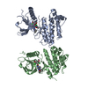
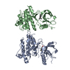
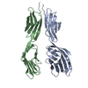
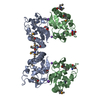
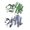
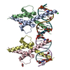
| Title | CRYSTAL STRUCTURE OF CLASS II RIBONUCLEASE H (RNASE HII) WITH MES LIGAND | ||||||
 Components Components | RIBONUCLEASE HII | ||||||
 Keywords Keywords |  HYDROLASE / Nuclease / Endonuclease / Structural Genomics / BSGC structure funded by NIH / Protein Structure Initiative / PSI / Berkeley Structural Genomics Center HYDROLASE / Nuclease / Endonuclease / Structural Genomics / BSGC structure funded by NIH / Protein Structure Initiative / PSI / Berkeley Structural Genomics Center | ||||||
| Function / homology |  Function and homology information Function and homology informationribonuclease H2 complex / DNA replication, removal of RNA primer / ribonuclease H / mismatch repair / RNA-DNA hybrid ribonuclease activity / manganese ion binding / RNA binding / cytoplasmSimilarity search - Function | ||||||
| Biological species |   Methanocaldococcus jannaschii (archaea) Methanocaldococcus jannaschii (archaea) | ||||||
| Method | X-RAY DIFFRACTION / SYNCHROTRON / MAD PHASING / Resolution: 2 Å | ||||||
 Authors Authors | Lai, L.H. / Yokota, H. / Hung, L.W. / Kim, R. / Kim, S.H. / Berkeley Structural Genomics Center (BSGC) | ||||||
 Citation Citation | Journal: Structure / Year: 2000 Title: Crystal structure of archaeal RNase HII: a homologue of human major RNase H Authors: Lai, L. / Yokota, H. / Hung, L.W. / Kim, R. / Kim, S.H. | ||||||
| History |
|
- Structure visualization
| Structure viewer | Molecule: MolmilJmol/JSmol |
|---|
- Downloads & links
-Download
| PDBx/mmCIF format | 1eke.cif.gz | 103.4 KB | Display | PDBx/mmCIF format |
|---|---|---|---|---|
| PDB format | pdb1eke.ent.gz | 84.7 KB | Display | PDB format |
| PDBx/mmJSON format | 1eke.json.gz | Tree view | PDBx/mmJSON format | |
| Others |  Other downloads Other downloads |
-Validation report
| Arichive directory | https://data.pdbj.org/pub/pdb/validation_reports/ek/1ekeftp://data.pdbj.org/pub/pdb/validation_reports/ek/1eke | HTTPS FTP |
|---|
-Related structure data
| Similar structure data | |
|---|---|
| Other databases |


-Links
 PDBj
PDBj
- Assembly
| Deposited unit | 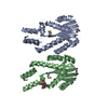
| ||||||||
|---|---|---|---|---|---|---|---|---|---|
| 1 |
| ||||||||
| Unit cell |
|
-Components
| #1: Protein | Mass: 26693.609 Da / Num. of mol.: 2 Source method: isolated from a genetically manipulated source Source: (gene. exp.) Methanocaldococcus jannaschii (archaea)Production host:  Escherichia coli (E. coli) / References: UniProt: Q57599, ribonuclease H Escherichia coli (E. coli) / References: UniProt: Q57599, ribonuclease H#2: Chemical | MES (buffer)  Mass: 195.237 Da / Num. of mol.: 2 / Source method: obtained synthetically / Formula: C6H13NO4S / Comment: pH buffer*YM Mass: 195.237 Da / Num. of mol.: 2 / Source method: obtained synthetically / Formula: C6H13NO4S / Comment: pH buffer*YM#3: Water | ChemComp-HOH / | Water Mass: 18.015 Da / Num. of mol.: 313 / Fragment: 2-(N-MORPHOLINO)ETHANESULFONATE / Source method: isolated from a natural source / Formula: H2O Mass: 18.015 Da / Num. of mol.: 313 / Fragment: 2-(N-MORPHOLINO)ETHANESULFONATE / Source method: isolated from a natural source / Formula: H2O |
|---|
-Experimental details
-Experiment
| Experiment | Method: X-RAY DIFFRACTION / Number of used crystals: 1 |
|---|
- Sample preparation
| Crystal | Density Matthews: 2.36 Å3/Da / Density % sol: 48 % | ||||||||||||||||||||||||||||||||||||||||||||||||||||||
|---|---|---|---|---|---|---|---|---|---|---|---|---|---|---|---|---|---|---|---|---|---|---|---|---|---|---|---|---|---|---|---|---|---|---|---|---|---|---|---|---|---|---|---|---|---|---|---|---|---|---|---|---|---|---|---|
| Crystal grow | Temperature: 293 K / Method: seeding / pH: 6.2 Details: PEG 3350, potassium thiocyanate, magnesium chloride, pH 6.2, seeding, temperature 293K | ||||||||||||||||||||||||||||||||||||||||||||||||||||||
| Crystal grow | *PLUS Temperature: 22 ℃ / Method: vapor diffusion, hanging drop / Details: used seeding | ||||||||||||||||||||||||||||||||||||||||||||||||||||||
| Components of the solutions | *PLUS
|
-Data collection
| Diffraction | Mean temperature: 120 K |
|---|---|
| Diffraction source | Source: SYNCHROTRON / Site: ALS  / Beamline: 5.0.2 / Wavelength: 1 / Beamline: 5.0.2 / Wavelength: 1 |
| Detector | Detector: CCD |
| Radiation | Protocol: MAD / Monochromatic (M) / Laue (L): M / Scattering type: x-ray |
| Radiation wavelength | Wavelength: 1 Å / Relative weight: 1 |
| Reflection | Resolution: 2→20 Å / Num. all: 30523 / Num. obs: 29715 / % possible obs: 97.4 % / Observed criterion σ(F): 0 / Observed criterion σ(I): 0 / Redundancy: 5.8 % / Biso Wilson estimate: 14.1 Å2 / Rmerge(I) obs: 0.059 / Net I/σ(I): 18.3 |
| Reflection shell | Resolution: 2→2.03 Å / Redundancy: 5.1 % / Rmerge(I) obs: 0.225 / Num. unique all: 1410 / % possible all: 94.6 |
| Reflection | *PLUS Num. measured all: 173694 |
| Reflection shell | *PLUS % possible obs: 94.6 % |
- Processing
| Software |
| |||||||||||||||||||||||||
|---|---|---|---|---|---|---|---|---|---|---|---|---|---|---|---|---|---|---|---|---|---|---|---|---|---|---|
| Refinement | Method to determine structure: MAD PHASING / Resolution: 2→20 Å / Rfactor Rfree error: 0.007 / Data cutoff high absF: 346794.7 / Data cutoff high rms absF: 10000 / Data cutoff low absF: 0 / Isotropic thermal model: RESTRAINED / Cross valid method: THROUGHOUT / σ(F): 0 / σ(I): 0 / Stereochemistry target values: cns / Details: Used Maximum likelihood target
| |||||||||||||||||||||||||
| Solvent computation | Solvent model: FLAT MODEL / Bsol: 45.92 Å2 / ksol: 0.325 e/Å3 | |||||||||||||||||||||||||
| Displacement parameters | Biso mean: 33.7 Å2
| |||||||||||||||||||||||||
| Refine analyze |
| |||||||||||||||||||||||||
| Refinement step | Cycle: LAST / Resolution: 2→20 Å
| |||||||||||||||||||||||||
| Refine LS restraints |
| |||||||||||||||||||||||||
| LS refinement shell | Resolution: 2→2.13 Å / Rfactor Rfree error: 0.021 / Total num. of bins used: 6
| |||||||||||||||||||||||||
| Xplor file |
| |||||||||||||||||||||||||
| Software | *PLUS Name: CNS / Version: 0.5 / Classification: refinement | |||||||||||||||||||||||||
| Refinement | *PLUS σ(F): 0 / % reflection Rfree: 4.9 % / Rfactor obs: 0.21 / Rfactor Rwork: 0.21 | |||||||||||||||||||||||||
| Solvent computation | *PLUS | |||||||||||||||||||||||||
| Displacement parameters | *PLUS Biso mean: 33.7 Å2 | |||||||||||||||||||||||||
| Refine LS restraints | *PLUS
| |||||||||||||||||||||||||
| LS refinement shell | *PLUS Rfactor Rfree: 0.306 / % reflection Rfree: 5 % / Rfactor Rwork: 0.25 |
