 Movie
Movie Controller

[English] 日本語
 Yorodumi
- PDB-4xhq: Re-refinement the crystal structure of Dscam1 isoform 1.34, N-ter... -
+ Open data

- Basic information
| Entry | Database: PDB / ID: 4xhq | |||||||||
|---|---|---|---|---|---|---|---|---|---|---|
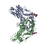
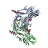
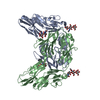
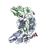
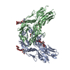
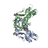
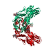
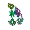
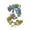
| Title | Re-refinement the crystal structure of Dscam1 isoform 1.34, N-terminal four Ig domains | |||||||||
 Components Components | Dscam | |||||||||
 Keywords Keywords | CELL ADHESION / Ig fold | |||||||||
| Function / homology |  Function and homology information Function and homology informationDSCAM interactions / mushroom body development / detection of mechanical stimulus involved in sensory perception of touch / detection of molecule of bacterial origin / central nervous system morphogenesis / ventral cord development / dendrite self-avoidance / cell-cell adhesion mediator activity / axon extension involved in axon guidance / axon guidance receptor activity ...DSCAM interactions / mushroom body development / detection of mechanical stimulus involved in sensory perception of touch / detection of molecule of bacterial origin / central nervous system morphogenesis / ventral cord development / dendrite self-avoidance / cell-cell adhesion mediator activity / axon extension involved in axon guidance / axon guidance receptor activity / peripheral nervous system development / axonal fasciculation / regulation of dendrite morphogenesis / regulation of axonogenesis / homophilic cell adhesion via plasma membrane adhesion molecules / neuron development / phagocytosis / antigen binding / axon guidance / perikaryon / neuron projection / axon / neuronal cell body / dendrite / protein homodimerization activity / extracellular region / identical protein binding / plasma membraneSimilarity search - Function | |||||||||
| Biological species |  Drosophila melanogaster (fruit fly) Drosophila melanogaster (fruit fly) | |||||||||
| Method | X-RAY DIFFRACTION / Resolution: 1.948 Å | |||||||||
 Authors Authors | Chen, Q. / Yu, Y. | |||||||||
 Citation Citation | Journal: NATURE / Year: 2007 Title: Structural Basis of Dscam Isoform Specificity Authors: Meijers, R. / Puettmann-Holgado, R. / Skiniotis, G. / Liu, J.-H. / Walz, T. / Wang, J.-H. / Schmucker, D. | |||||||||
| History |
| |||||||||
| Remark 0 | THIS ENTRY 4XHQ REFLECTS AN ALTERNATIVE MODELING OF THE STRUCTURAL DATA IN R2V5MSF ORIGINAL DATA ...THIS ENTRY 4XHQ REFLECTS AN ALTERNATIVE MODELING OF THE STRUCTURAL DATA IN R2V5MSF ORIGINAL DATA DETERMINED BY AUTHOR: R.MEIJERS,R.PUETTMANN-HOLGADO,G.SKINIOTIS,J.-H.LIU,T.WALZ, D.SCHMUCKER,J.-H.WANG |
- Structure visualization
| Structure viewer | Molecule: MolmilJmol/JSmol |
|---|
- Downloads & links
-Download
| PDBx/mmCIF format | 4xhq.cif.gz | 100.7 KB | Display | PDBx/mmCIF format |
|---|---|---|---|---|
| PDB format | pdb4xhq.ent.gz | 78.8 KB | Display | PDB format |
| PDBx/mmJSON format | 4xhq.json.gz | Tree view | PDBx/mmJSON format | |
| Others |  Other downloads Other downloads |
-Validation report
| Arichive directory | https://data.pdbj.org/pub/pdb/validation_reports/xh/4xhqftp://data.pdbj.org/pub/pdb/validation_reports/xh/4xhq | HTTPS FTP |
|---|
-Related structure data
| Related structure data |  4wvrC 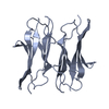 4x5lC  4x83C  4x8xC  4x9bC 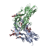 4x9fC  4x9gC 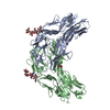 4x9hC 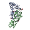 4x9iC  4xb7C  4xb8C C: citing same article ( |
|---|---|
| Similar structure data |


-Links
 PDBj
PDBj


- Assembly
| Deposited unit | 
| |||||||||
|---|---|---|---|---|---|---|---|---|---|---|
| 1 | 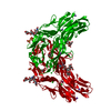
| |||||||||
| Unit cell |
| |||||||||
| Components on special symmetry positions |
|
-Components
-Protein , 1 types, 1 molecules A
| #1: Protein | Mass: 42880.582 Da / Num. of mol.: 1 / Fragment: UNP residues 36-423 Source method: isolated from a genetically manipulated source Source: (gene. exp.) Drosophila melanogaster (fruit fly) / Gene: Dscam1, Dscam, CG17800 / Production host:  Spodoptera frugiperda (fall armyworm) / References: UniProt: Q9NBA1 Spodoptera frugiperda (fall armyworm) / References: UniProt: Q9NBA1 |
|---|
-Sugars , 2 types, 2 molecules
| #2: Polysaccharide | beta-D-mannopyranose-(1-4)-2-acetamido-2-deoxy-beta-D-glucopyranose-(1-4)-2-acetamido-2-deoxy-beta- ...beta-D-mannopyranose-(1-4)-2-acetamido-2-deoxy-beta-D-glucopyranose-(1-4)-2-acetamido-2-deoxy-beta-D-glucopyranose / Mass: 586.542 Da / Num. of mol.: 1 Source method: isolated from a genetically manipulated source |
|---|---|
| #3: Polysaccharide | alpha-D-mannopyranose-(1-3)-[alpha-D-mannopyranose-(1-6)]beta-D-mannopyranose-(1-4)-2-acetamido-2- ...alpha-D-mannopyranose-(1-3)-[alpha-D-mannopyranose-(1-6)]beta-D-mannopyranose-(1-4)-2-acetamido-2-deoxy-beta-D-glucopyranose-(1-4)-2-acetamido-2-deoxy-beta-D-glucopyranose / Mass: 910.823 Da / Num. of mol.: 1 Source method: isolated from a genetically manipulated source |
-Non-polymers , 5 types, 396 molecules 


| #4: Chemical | Glycerol Mass: 92.094 Da / Num. of mol.: 3 / Source method: obtained synthetically / Formula: C3H8O3 Mass: 92.094 Da / Num. of mol.: 3 / Source method: obtained synthetically / Formula: C3H8O3#5: Chemical | ChemComp-CL / Chloride Mass: 35.453 Da / Num. of mol.: 4 / Source method: obtained synthetically / Formula: Cl Mass: 35.453 Da / Num. of mol.: 4 / Source method: obtained synthetically / Formula: Cl#6: Chemical | ChemComp-NA /  Mass: 22.990 Da / Num. of mol.: 4 / Source method: obtained synthetically / Formula: Na Mass: 22.990 Da / Num. of mol.: 4 / Source method: obtained synthetically / Formula: Na#7: Chemical | Sulfate Mass: 96.063 Da / Num. of mol.: 2 / Source method: obtained synthetically / Formula: SO4 Mass: 96.063 Da / Num. of mol.: 2 / Source method: obtained synthetically / Formula: SO4#8: Water | ChemComp-HOH / | WaterMass: 18.015 Da / Num. of mol.: 383 / Source method: isolated from a natural source / Formula: H2O |
|---|
-Experimental details
-Experiment
| Experiment | Method: X-RAY DIFFRACTION |
|---|
- Sample preparation
| Crystal | Density Matthews: 4.7 Å3/Da / Density % sol: 73.84 % / Description: AUTHOR USED THE SF DATA FROM ENTRY 2V5M. |
|---|---|
| Crystal grow | Details: 1.5 M AMMONIUM SULPHATE, 0.1 M HEPES PH 7.5 |
-Data collection
| Radiation | Scattering type: x-ray |
|---|---|
| Radiation wavelength | Relative weight: 1 |
- Processing
| Software |
| ||||||||||||||||||||||||||||||||||||||||||||||||||||||||||||||||||||||||||||||||||||||||||||||||||||||||||||||||||||||||||||||||||||||||||||||||||||||||||
|---|---|---|---|---|---|---|---|---|---|---|---|---|---|---|---|---|---|---|---|---|---|---|---|---|---|---|---|---|---|---|---|---|---|---|---|---|---|---|---|---|---|---|---|---|---|---|---|---|---|---|---|---|---|---|---|---|---|---|---|---|---|---|---|---|---|---|---|---|---|---|---|---|---|---|---|---|---|---|---|---|---|---|---|---|---|---|---|---|---|---|---|---|---|---|---|---|---|---|---|---|---|---|---|---|---|---|---|---|---|---|---|---|---|---|---|---|---|---|---|---|---|---|---|---|---|---|---|---|---|---|---|---|---|---|---|---|---|---|---|---|---|---|---|---|---|---|---|---|---|---|---|---|---|---|---|
| Refinement | Resolution: 1.948→19.834 Å / SU ML: 0.25 / Cross valid method: FREE R-VALUE / σ(F): 1.34 / Phase error: 26.5 / Stereochemistry target values: ML
| ||||||||||||||||||||||||||||||||||||||||||||||||||||||||||||||||||||||||||||||||||||||||||||||||||||||||||||||||||||||||||||||||||||||||||||||||||||||||||
| Solvent computation | Shrinkage radii: 0.9 Å / VDW probe radii: 1.11 Å / Solvent model: FLAT BULK SOLVENT MODEL | ||||||||||||||||||||||||||||||||||||||||||||||||||||||||||||||||||||||||||||||||||||||||||||||||||||||||||||||||||||||||||||||||||||||||||||||||||||||||||
| Refinement step | Cycle: LAST / Resolution: 1.948→19.834 Å
| ||||||||||||||||||||||||||||||||||||||||||||||||||||||||||||||||||||||||||||||||||||||||||||||||||||||||||||||||||||||||||||||||||||||||||||||||||||||||||
| Refine LS restraints |
| ||||||||||||||||||||||||||||||||||||||||||||||||||||||||||||||||||||||||||||||||||||||||||||||||||||||||||||||||||||||||||||||||||||||||||||||||||||||||||
| LS refinement shell |
|
