 Movie
Movie Controller

[English] 日本語
 Yorodumi
- PDB-4x9h: Crystal structure of Dscam1 isoform 8.4, N-terminal four Ig domains -
+ Open data

- Basic information
| Entry | Database: PDB / ID: 4x9h | |||||||||
|---|---|---|---|---|---|---|---|---|---|---|
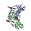
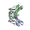
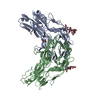
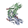
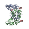
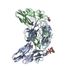
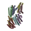
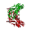
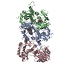
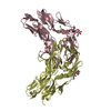
| Title | Crystal structure of Dscam1 isoform 8.4, N-terminal four Ig domains | |||||||||
 Components Components | Down syndrome cell adhesion molecule, isoform AP | |||||||||
 Keywords Keywords |  CELL ADHESION / Ig fold CELL ADHESION / Ig fold | |||||||||
| Function / homology |  Function and homology information Function and homology informationmushroom body development / detection of mechanical stimulus involved in sensory perception of touch / detection of molecule of bacterial origin / central nervous system morphogenesis / ventral cord development / dendrite self-avoidance / cell-cell adhesion mediator activity / axon extension involved in axon guidance / axon guidance receptor activity / peripheral nervous system development ...mushroom body development / detection of mechanical stimulus involved in sensory perception of touch / detection of molecule of bacterial origin / central nervous system morphogenesis / ventral cord development / dendrite self-avoidance / cell-cell adhesion mediator activity / axon extension involved in axon guidance / axon guidance receptor activity / peripheral nervous system development / axonal fasciculation / regulation of dendrite morphogenesis / regulation of axonogenesis / homophilic cell adhesion via plasma membrane adhesion molecules / neuron development / phagocytosis / antigen binding / axon guidance / neuron projection / axon / neuronal cell body / dendrite / protein homodimerization activity / identical protein binding / plasma membraneSimilarity search - Function | |||||||||
| Biological species |  Drosophila melanogaster (fruit fly) Drosophila melanogaster (fruit fly) | |||||||||
| Method | X-RAY DIFFRACTION / SYNCHROTRON / MOLECULAR REPLACEMENT / Resolution: 2.95 Å | |||||||||
 Authors Authors | Chen, Q. | |||||||||
 Citation Citation | Journal: Sci Adv / Year: 2016 Title: Structural basis of Dscam1 homodimerization: Insights into context constraint for protein recognition Authors: Li, S.A. / Cheng, L. / Yu, Y. / Chen, Q. | |||||||||
| History |
|
- Structure visualization
| Structure viewer | Molecule: MolmilJmol/JSmol |
|---|
- Downloads & links
-Download
| PDBx/mmCIF format | 4x9h.cif.gz | 167.9 KB | Display | PDBx/mmCIF format |
|---|---|---|---|---|
| PDB format | pdb4x9h.ent.gz | 131 KB | Display | PDB format |
| PDBx/mmJSON format | 4x9h.json.gz | Tree view | PDBx/mmJSON format | |
| Others |  Other downloads Other downloads |
-Validation report
| Arichive directory | https://data.pdbj.org/pub/pdb/validation_reports/x9/4x9hftp://data.pdbj.org/pub/pdb/validation_reports/x9/4x9h | HTTPS FTP |
|---|
-Related structure data
| Related structure data |  4wvrC 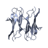 4x5lC  4x83C  4x8xC  4x9bC 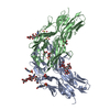 4x9fC  4x9gC 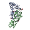 4x9iC  4xb7C  4xb8C  4xhqC 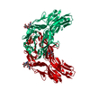 2v5mS C: citing same article ( S: Starting model for refinement |
|---|---|
| Similar structure data |
-Links
 PDBj
PDBj


- Assembly
| Deposited unit | 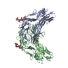
| ||||||||
|---|---|---|---|---|---|---|---|---|---|
| 1 |
| ||||||||
| Unit cell |
|
-Components
| #1: Protein | Mass: 44010.734 Da / Num. of mol.: 2 / Fragment: UNP residues 34-431 Source method: isolated from a genetically manipulated source Source: (gene. exp.) Drosophila melanogaster (fruit fly) / Gene: Dscam1, Dscam, CG17800, Dmel_CG17800 / Production host: Trichoplusia ni (cabbage looper) / References: UniProt: Q0E9L0#2: Polysaccharide | / Mass: 748.682 Da / Num. of mol.: 2 / Source method: obtained synthetically#3: Sugar | N-Acetylglucosamine  Type: D-saccharide, beta linking / Mass: 221.208 Da / Num. of mol.: 2 / Source method: obtained synthetically / Formula: C8H15NO6 Type: D-saccharide, beta linking / Mass: 221.208 Da / Num. of mol.: 2 / Source method: obtained synthetically / Formula: C8H15NO6#4: Water | ChemComp-HOH / | Water Mass: 18.015 Da / Num. of mol.: 3 / Source method: isolated from a natural source / Formula: H2O Mass: 18.015 Da / Num. of mol.: 3 / Source method: isolated from a natural source / Formula: H2O |
|---|
-Experimental details
-Experiment
| Experiment | Method: X-RAY DIFFRACTION / Number of used crystals: 1 |
|---|
- Sample preparation
| Crystal | Density Matthews: 3.04 Å3/Da / Density % sol: 59.52 % |
|---|---|
| Crystal grow | Temperature: 289 K / Method: vapor diffusion, hanging drop / Details: 0.1 M MES pH6.5, 20% (w/v) PEG 3350 |
-Data collection
| Diffraction | Mean temperature: 100 K |
|---|---|
| Diffraction source | Source: SYNCHROTRON / Site: APS  / Beamline: 19-ID / Wavelength: 1 Å / Beamline: 19-ID / Wavelength: 1 Å |
| Detector | Type: DECTRIS PILATUS 2M / Detector: PIXEL / Date: Jul 12, 2014 |
| Radiation | Monochromator: SiIII double crystal / Protocol: SINGLE WAVELENGTH / Monochromatic (M) / Laue (L): M / Scattering type: x-ray |
| Radiation wavelength | Wavelength: 1 Å / Relative weight: 1 |
| Reflection | Resolution: 2.95→50 Å / Num. obs: 22498 / % possible obs: 97.2 % / Redundancy: 2.7 % / Net I/σ(I): 14.1 |
- Processing
| Software |
| |||||||||||||||||||||||||||||||||||||||||||||||||||||||||||||||
|---|---|---|---|---|---|---|---|---|---|---|---|---|---|---|---|---|---|---|---|---|---|---|---|---|---|---|---|---|---|---|---|---|---|---|---|---|---|---|---|---|---|---|---|---|---|---|---|---|---|---|---|---|---|---|---|---|---|---|---|---|---|---|---|---|
| Refinement | Method to determine structure: MOLECULAR REPLACEMENT Starting model: 2V5M Resolution: 2.95→47.519 Å / SU ML: 0.57 / Cross valid method: FREE R-VALUE / σ(F): 1.34 / Phase error: 36.91 / Stereochemistry target values: ML
| |||||||||||||||||||||||||||||||||||||||||||||||||||||||||||||||
| Solvent computation | Shrinkage radii: 0.9 Å / VDW probe radii: 1.11 Å / Solvent model: FLAT BULK SOLVENT MODEL | |||||||||||||||||||||||||||||||||||||||||||||||||||||||||||||||
| Refinement step | Cycle: LAST / Resolution: 2.95→47.519 Å
| |||||||||||||||||||||||||||||||||||||||||||||||||||||||||||||||
| Refine LS restraints |
| |||||||||||||||||||||||||||||||||||||||||||||||||||||||||||||||
| LS refinement shell |
|
