 Movie
Movie Controller


+ Open data

- Basic information
| Entry | Database: PDB / ID: 6qyc | |||||||||
|---|---|---|---|---|---|---|---|---|---|---|
| Title | Crystal structure of MtrC from Shewanella baltica OS185 | |||||||||
 Components Components | Decaheme c-type cytochrome, OmcA/MtrC family | |||||||||
 Keywords Keywords |  ELECTRON TRANSPORT / Cytochrome / Membrane protein / greek key / multiheme ELECTRON TRANSPORT / Cytochrome / Membrane protein / greek key / multiheme | |||||||||
| Function / homology | Decahaem cytochrome, c-type, OmcA/MtrC / Multiheme cytochrome superfamily / HEME C / Multiheme cytochrome MtrC Function and homology information Function and homology information | |||||||||
| Biological species |  Shewanella baltica OS185 (bacteria) Shewanella baltica OS185 (bacteria) | |||||||||
| Method | X-RAY DIFFRACTION / SYNCHROTRON / SAD / Resolution: 2.29 Å | |||||||||
 Authors Authors | Clarke, T.A. / Edwards, M.J. | |||||||||
| Funding support |  United Kingdom, 2items United Kingdom, 2items
| |||||||||
 Citation Citation | Journal: Cell / Year: 2020 Title: The Crystal Structure of a Biological Insulated Transmembrane Molecular Wire. Authors: Edwards, M.J. / White, G.F. / Butt, J.N. / Richardson, D.J. / Clarke, T.A. | |||||||||
| History |
|
- Structure visualization
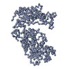
| Structure viewer | Molecule: MolmilJmol/JSmol |
|---|
- Downloads & links
-Download
| PDBx/mmCIF format | 6qyc.cif.gz | 398.4 KB | Display | PDBx/mmCIF format |
|---|---|---|---|---|
| PDB format | pdb6qyc.ent.gz | 338.9 KB | Display | PDB format |
| PDBx/mmJSON format | 6qyc.json.gz | Tree view | PDBx/mmJSON format | |
| Others |  Other downloads Other downloads |
-Validation report
| Arichive directory | https://data.pdbj.org/pub/pdb/validation_reports/qy/6qycftp://data.pdbj.org/pub/pdb/validation_reports/qy/6qyc | HTTPS FTP |
|---|
-Related structure data


-Links
 PDBj
PDBj


- Assembly
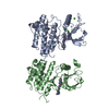
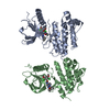
| Deposited unit | 
| ||||||||
|---|---|---|---|---|---|---|---|---|---|
| 1 | 
| ||||||||
| 2 | 
| ||||||||
| 3 | 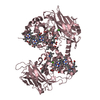
| ||||||||
| Unit cell |
|
-Components
| #1: Protein | Mass: 64129.344 Da / Num. of mol.: 3 Source method: isolated from a genetically manipulated source Source: (gene. exp.) Shewanella baltica OS185 (bacteria) / Gene: NCTC10735_02683 / Production host: Shewanella oneidensis MR-1 (bacteria) / Strain (production host): MR-1 / References: UniProt: P0DSN4*PLUS#2: Chemical | ChemComp-HEC / Heme C  Mass: 618.503 Da / Num. of mol.: 30 / Source method: obtained synthetically / Formula: C34H34FeN4O4 Mass: 618.503 Da / Num. of mol.: 30 / Source method: obtained synthetically / Formula: C34H34FeN4O4#3: Chemical | ChemComp-CA /   Mass: 40.078 Da / Num. of mol.: 8 / Source method: obtained synthetically / Formula: Ca Mass: 40.078 Da / Num. of mol.: 8 / Source method: obtained synthetically / Formula: Ca#4: Chemical | Ethylene glycol  Mass: 62.068 Da / Num. of mol.: 2 / Source method: obtained synthetically / Formula: C2H6O2 Mass: 62.068 Da / Num. of mol.: 2 / Source method: obtained synthetically / Formula: C2H6O2#5: Water | ChemComp-HOH / | Water Mass: 18.015 Da / Num. of mol.: 913 / Source method: isolated from a natural source / Formula: H2O Mass: 18.015 Da / Num. of mol.: 913 / Source method: isolated from a natural source / Formula: H2O |
|---|
-Experimental details
-Experiment
| Experiment | Method: X-RAY DIFFRACTION / Number of used crystals: 1 |
|---|
- Sample preparation
| Crystal | Density Matthews: 3 Å3/Da / Density % sol: 58.95 % |
|---|---|
| Crystal grow | Temperature: 289 K / Method: vapor diffusion, sitting drop / pH: 4.5 Details: 0.4 M Sodium Acetate, 5% PEG 6000, 5% PEG 8000, 5% PEG 10000 |
-Data collection
| Diffraction | Mean temperature: 100 K / Serial crystal experiment: N |
|---|---|
| Diffraction source | Source: SYNCHROTRON / Site: Diamond / Beamline: I04 / Wavelength: 1.72001 Å |
| Detector | Type: DECTRIS PILATUS 2M / Detector: PIXEL / Date: May 7, 2018 |
| Radiation | Protocol: SINGLE WAVELENGTH / Monochromatic (M) / Laue (L): M / Scattering type: x-ray |
| Radiation wavelength | Wavelength: 1.72001 Å / Relative weight: 1 |
| Reflection | Resolution: 2.29→90.52 Å / Num. obs: 104279 / % possible obs: 99.5 % / Redundancy: 12.6 % / Biso Wilson estimate: 50.79 Å2 / CC1/2: 0.999 / Rmerge(I) obs: 0.112 / Rpim(I) all: 0.033 / Rrim(I) all: 0.117 / Net I/σ(I): 17 / Num. measured all: 1317009 / Scaling rejects: 3 |
| Reflection shell | Resolution: 2.29→2.35 Å / Redundancy: 10.2 % / Rmerge(I) obs: 1.887 / Num. unique obs: 7485 / CC1/2: 0.475 / Rpim(I) all: 0.612 / Rrim(I) all: 1.986 / % possible all: 98.2 |
- Processing
| Software |
| |||||||||||||||||||||||||||||||||||||||||||||||||||||||||||||||||||||||||||||||||||||||||||||||||||||||||||||||||||||||||||||||||||||||||||||||||||||||||||||||||||||||||||||||||||||||||||||||||||||||||||||||||||||||||
|---|---|---|---|---|---|---|---|---|---|---|---|---|---|---|---|---|---|---|---|---|---|---|---|---|---|---|---|---|---|---|---|---|---|---|---|---|---|---|---|---|---|---|---|---|---|---|---|---|---|---|---|---|---|---|---|---|---|---|---|---|---|---|---|---|---|---|---|---|---|---|---|---|---|---|---|---|---|---|---|---|---|---|---|---|---|---|---|---|---|---|---|---|---|---|---|---|---|---|---|---|---|---|---|---|---|---|---|---|---|---|---|---|---|---|---|---|---|---|---|---|---|---|---|---|---|---|---|---|---|---|---|---|---|---|---|---|---|---|---|---|---|---|---|---|---|---|---|---|---|---|---|---|---|---|---|---|---|---|---|---|---|---|---|---|---|---|---|---|---|---|---|---|---|---|---|---|---|---|---|---|---|---|---|---|---|---|---|---|---|---|---|---|---|---|---|---|---|---|---|---|---|---|---|---|---|---|---|---|---|---|---|---|---|---|---|---|---|---|
| Refinement | Method to determine structure: SAD / Resolution: 2.29→87.2 Å / SU ML: 0.32 / Cross valid method: THROUGHOUT / σ(F): 1.34 / Phase error: 24.66
| |||||||||||||||||||||||||||||||||||||||||||||||||||||||||||||||||||||||||||||||||||||||||||||||||||||||||||||||||||||||||||||||||||||||||||||||||||||||||||||||||||||||||||||||||||||||||||||||||||||||||||||||||||||||||
| Solvent computation | Shrinkage radii: 0.9 Å / VDW probe radii: 1.11 Å | |||||||||||||||||||||||||||||||||||||||||||||||||||||||||||||||||||||||||||||||||||||||||||||||||||||||||||||||||||||||||||||||||||||||||||||||||||||||||||||||||||||||||||||||||||||||||||||||||||||||||||||||||||||||||
| Displacement parameters | Biso max: 117.89 Å2 / Biso mean: 52.3964 Å2 / Biso min: 30.4 Å2 | |||||||||||||||||||||||||||||||||||||||||||||||||||||||||||||||||||||||||||||||||||||||||||||||||||||||||||||||||||||||||||||||||||||||||||||||||||||||||||||||||||||||||||||||||||||||||||||||||||||||||||||||||||||||||
| Refinement step | Cycle: final / Resolution: 2.29→87.2 Å
| |||||||||||||||||||||||||||||||||||||||||||||||||||||||||||||||||||||||||||||||||||||||||||||||||||||||||||||||||||||||||||||||||||||||||||||||||||||||||||||||||||||||||||||||||||||||||||||||||||||||||||||||||||||||||
| LS refinement shell | Refine-ID: X-RAY DIFFRACTION / Rfactor Rfree error: 0 / Total num. of bins used: 30
|
