- PDB-2qm2: Putative HopJ type III effector protein from Vibrio parahaemolyticus -
+
Open data
ID or keywords:
Loading...
-
Basic information
Entry
Database: PDB / ID: 2qm2
Title
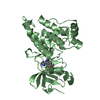
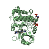
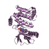
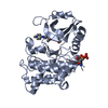
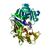
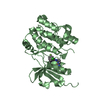
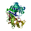
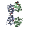
Putative HopJ type III effector protein from Vibrio parahaemolyticus
Components
Putative HopJ type III effector protein
Keywords
STRUCTURAL GENOMICS / UNKNOWN FUNCTION / alpha-beta structure / PSI-2 / Protein Structure Initiative / Midwest Center for Structural Genomics / MCSG
Function / homology
pseudo Tubby roll / vpa0580 domain like / HopJ type III effector protein / Type III effector HopJ superfamily / HopJ type III effector protein / Alpha-Beta Barrel / Alpha Beta / : / Type III effector
Function and homology information
Biological species
Vibrio parahaemolyticus (bacteria)
Method
X-RAY DIFFRACTION / SYNCHROTRON / MOLECULAR REPLACEMENT, SAD / Resolution: 2.09 Å
BIOMOLECULE: 1 AUTHORS STATE THAT THE BIOLOGICAL UNIT OF THIS PROTEIN IS UNKNOWN. SEE REMARK 350 ... BIOMOLECULE: 1 AUTHORS STATE THAT THE BIOLOGICAL UNIT OF THIS PROTEIN IS UNKNOWN. SEE REMARK 350 FOR THE PROGRAM GENERATED ASSEMBLY INFORMATION FOR THE STRUCTURE IN THIS ENTRY. THE REMARK MAY ALSO PROVIDE INFORMATION ON BURIED SURFACE AREA.
In the structure databanks used in Yorodumi, some data are registered as the other names, "COVID-19 virus" and "2019-nCoV". Here are the details of the virus and the list of structure data.
Jan 31, 2019. EMDB accession codes are about to change! (news from PDBe EMDB page)
EMDB accession codes are about to change! (news from PDBe EMDB page)
The allocation of 4 digits for EMDB accession codes will soon come to an end. Whilst these codes will remain in use, new EMDB accession codes will include an additional digit and will expand incrementally as the available range of codes is exhausted. The current 4-digit format prefixed with “EMD-” (i.e. EMD-XXXX) will advance to a 5-digit format (i.e. EMD-XXXXX), and so on. It is currently estimated that the 4-digit codes will be depleted around Spring 2019, at which point the 5-digit format will come into force.
The EM Navigator/Yorodumi systems omit the EMD- prefix.
Related info.:Q: What is EMD? / ID/Accession-code notation in Yorodumi/EM Navigator
Yorodumi is a browser for structure data from EMDB, PDB, SASBDB, etc.
This page is also the successor to EM Navigator detail page, and also detail information page/front-end page for Omokage search.
The word "yorodu" (or yorozu) is an old Japanese word meaning "ten thousand". "mi" (miru) is to see.
Related info.:EMDB / PDB / SASBDB / Comparison of 3 databanks / Yorodumi Search / Aug 31, 2016. New EM Navigator & Yorodumi / Yorodumi Papers / Jmol/JSmol / Function and homology information / Changes in new EM Navigator and Yorodumi
 Movie
Movie Controller

 Yorodumi
 Open data
 Basic information
 Components
 Keywords
 STRUCTURAL GENOMICS / UNKNOWN FUNCTION / alpha-beta structure / PSI-2 /
  Function and homology information

 Authors
 Citation
 Structure visualization
 Downloads & links
 Other downloads


 PDBj
PDBj Assembly


 Mass: 39.098 Da / Num. of mol.: 1 / Source method: obtained synthetically / Formula: K
 Mass: 92.094 Da / Num. of mol.: 1 / Source method: obtained synthetically / Formula: C3H8O3
 Mass: 22.990 Da / Num. of mol.: 1 / Source method: obtained synthetically / Formula: Na
 Mass: 122.143 Da / Num. of mol.: 1 / Source method: obtained synthetically / Formula: C4H12NO3 / Comment: pH buffer*YM
 Sample preparation
 / Beamline: 19-ID / Wavelength: 0.9793 Å
 Processing