 Movie
Movie Controller

[English] 日本語
 Yorodumi
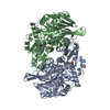
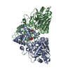
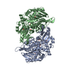
- PDB-1ecj: ESCHERICHIA COLI GLUTAMINE PHOSPHORIBOSYLPYROPHOSPHATE (PRPP) AMI... -
+ Open data

- Basic information
| Entry | Database: PDB / ID: 1ecj | ||||||
|---|---|---|---|---|---|---|---|
| Title | ESCHERICHIA COLI GLUTAMINE PHOSPHORIBOSYLPYROPHOSPHATE (PRPP) AMIDOTRANSFERASE COMPLEXED WITH 2 AMP PER TETRAMER | ||||||
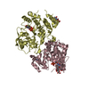
 Components Components | GLUTAMINE PHOSPHORIBOSYLPYROPHOSPHATE AMIDOTRANSFERASE | ||||||
 Keywords Keywords |  TRANSFERASE / GLUTAMINE AMIDOTRANSFERASE / PURINE BIOSYNTHESIS / GLYCOSYLTRANSFERASE / AMP / ADENINE 5'-MONOPHOSPHATE TRANSFERASE / GLUTAMINE AMIDOTRANSFERASE / PURINE BIOSYNTHESIS / GLYCOSYLTRANSFERASE / AMP / ADENINE 5'-MONOPHOSPHATE | ||||||
| Function / homology |  Function and homology informationamidophosphoribosyltransferase / amidophosphoribosyltransferase activity / purine nucleobase biosynthetic process / purine nucleotide biosynthetic process / 'de novo' IMP biosynthetic process / guanosine tetraphosphate binding / glutamine metabolic process / glycosyltransferase activity / magnesium ion binding / identical protein binding ...amidophosphoribosyltransferase / amidophosphoribosyltransferase activity / purine nucleobase biosynthetic process / purine nucleotide biosynthetic process / 'de novo' IMP biosynthetic process / guanosine tetraphosphate binding / glutamine metabolic process / glycosyltransferase activity / magnesium ion binding / identical protein binding / cytosol / cytoplasm Function and homology informationamidophosphoribosyltransferase / amidophosphoribosyltransferase activity / purine nucleobase biosynthetic process / purine nucleotide biosynthetic process / 'de novo' IMP biosynthetic process / guanosine tetraphosphate binding / glutamine metabolic process / glycosyltransferase activity / magnesium ion binding / identical protein binding ...amidophosphoribosyltransferase / amidophosphoribosyltransferase activity / purine nucleobase biosynthetic process / purine nucleotide biosynthetic process / 'de novo' IMP biosynthetic process / guanosine tetraphosphate binding / glutamine metabolic process / glycosyltransferase activity / magnesium ion binding / identical protein binding / cytosol / cytoplasmSimilarity search - Function | ||||||
| Biological species |  Escherichia coli (E. coli) Escherichia coli (E. coli) | ||||||
| Method | X-RAY DIFFRACTION / SYNCHROTRON / MOLECULAR REPLACEMENT / Resolution: 2.5 Å | ||||||
 Authors Authors | Muchmore, C.R. / Krahn, J.M. / Smith, J.L. | ||||||
 Citation Citation | Journal: Protein Sci. / Year: 1998 Title: Crystal structure of glutamine phosphoribosylpyrophosphate amidotransferase from Escherichia coli. Authors: Muchmore, C.R. / Krahn, J.M. / Kim, J.H. / Zalkin, H. / Smith, J.L. | ||||||
| History |
|
- Structure visualization
| Structure viewer | Molecule: MolmilJmol/JSmol |
|---|
- Downloads & links
-Download
| PDBx/mmCIF format | 1ecj.cif.gz | 382.5 KB | Display | PDBx/mmCIF format |
|---|---|---|---|---|
| PDB format | pdb1ecj.ent.gz | 316 KB | Display | PDB format |
| PDBx/mmJSON format | 1ecj.json.gz | Tree view | PDBx/mmJSON format | |
| Others |  Other downloads Other downloads |
-Validation report
| Arichive directory | https://data.pdbj.org/pub/pdb/validation_reports/ec/1ecjftp://data.pdbj.org/pub/pdb/validation_reports/ec/1ecj | HTTPS FTP |
|---|
-Related structure data
| Related structure data |  1ecfC 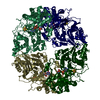 1gphS S: Starting model for refinement C: citing same article ( |
|---|---|
| Similar structure data |


-Links
 PDBj
PDBj


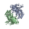
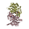
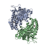
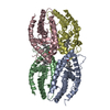
- Assembly
| Deposited unit | 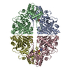
| ||||||||||||||||
|---|---|---|---|---|---|---|---|---|---|---|---|---|---|---|---|---|---|
| 1 | 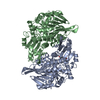
| ||||||||||||||||
| 2 | 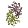
| ||||||||||||||||
| 3 |
| ||||||||||||||||
| Unit cell |
| ||||||||||||||||
| Noncrystallographic symmetry (NCS) | NCS oper:
|
-Components
| #1: Protein | Mass: 56422.684 Da / Num. of mol.: 4 Source method: isolated from a genetically manipulated source Source: (gene. exp.) Escherichia coli (E. coli) / Gene: PURF / Plasmid: PT7F1 / Species (production host): Escherichia coli / Production host: Escherichia coli BL21(DE3) (bacteria) / Strain (production host): BL21 (DE3)References: UniProt: P00496, UniProt: P0AG16*PLUS, amidophosphoribosyltransferase#2: Chemical | Adenosine monophosphate  Mass: 347.221 Da / Num. of mol.: 2 / Source method: obtained synthetically / Formula: C10H14N5O7P / Comment: AMP*YM Mass: 347.221 Da / Num. of mol.: 2 / Source method: obtained synthetically / Formula: C10H14N5O7P / Comment: AMP*YM#3: Water | ChemComp-HOH / | Water Mass: 18.015 Da / Num. of mol.: 178 / Source method: isolated from a natural source / Formula: H2O Mass: 18.015 Da / Num. of mol.: 178 / Source method: isolated from a natural source / Formula: H2O |
|---|
-Experimental details
-Experiment
| Experiment | Method: X-RAY DIFFRACTION / Number of used crystals: 10 |
|---|
- Sample preparation
| Crystal | Density Matthews: 2.37 Å3/Da / Density % sol: 47.6 % Description: THE SYNCHROTRON DATA DESCRIBED BY THE PREVIOUS STATISTICS WAS SUPPLEMENTED WITH LOW RESOLUTION DATA COLLECTED WITH A RIGAKU ROTATING ANODE COPPER K(ALPHA) SOURCE AND A XUONG-HAMLIN ...Description: THE SYNCHROTRON DATA DESCRIBED BY THE PREVIOUS STATISTICS WAS SUPPLEMENTED WITH LOW RESOLUTION DATA COLLECTED WITH A RIGAKU ROTATING ANODE COPPER K(ALPHA) SOURCE AND A XUONG-HAMLIN MULTIWIRE AREA DETECTOR. THIS DATA WAS PROCESSED WITH XDS (KABSCH) ADAPTED FOR THE XUONG-HAMLIN DETECTOR (D. DIGGS, K. WATKINS). | ||||||||||||||||||||||||||||||||||||||||||||||||||||||||||||
|---|---|---|---|---|---|---|---|---|---|---|---|---|---|---|---|---|---|---|---|---|---|---|---|---|---|---|---|---|---|---|---|---|---|---|---|---|---|---|---|---|---|---|---|---|---|---|---|---|---|---|---|---|---|---|---|---|---|---|---|---|---|
| Crystal grow | pH: 6.3 Details: 100MM MES, 50MM TRIS, 2MM AMP, 4MM EDTA, 15-18% PEG-3350, PH 6.3 | ||||||||||||||||||||||||||||||||||||||||||||||||||||||||||||
| Crystal grow | *PLUS Temperature: 27 ℃ / Method: vapor diffusion, sitting drop | ||||||||||||||||||||||||||||||||||||||||||||||||||||||||||||
| Components of the solutions | *PLUS
|
-Data collection
| Diffraction | Mean temperature: 279 K |
|---|---|
| Diffraction source | Source: SYNCHROTRON / Site: Photon Factory  / Beamline: BL-6A / Wavelength: 1 / Beamline: BL-6A / Wavelength: 1 |
| Detector | Type: FUJI / Detector: IMAGE PLATE / Date: Feb 1, 1992 |
| Radiation | Monochromatic (M) / Laue (L): M / Scattering type: x-ray |
| Radiation wavelength | Wavelength: 1 Å / Relative weight: 1 |
| Reflection | Resolution: 2.5→30 Å / Num. obs: 55291 / % possible obs: 77.8 % / Redundancy: 3.6 % / Rsym value: 0.065 / Net I/σ(I): 37 |
| Reflection shell | Resolution: 2.5→2.61 Å / Redundancy: 1.9 % / Mean I/σ(I) obs: 4.3 / Rsym value: 0.215 / % possible all: 56.1 |
| Reflection | *PLUS Num. measured all: 199847 / Rmerge(I) obs: 0.065 |
- Processing
| Software |
| ||||||||||||||||||||||||||||||||||||||||||||||||||||||||||||||||||||||||||||||||
|---|---|---|---|---|---|---|---|---|---|---|---|---|---|---|---|---|---|---|---|---|---|---|---|---|---|---|---|---|---|---|---|---|---|---|---|---|---|---|---|---|---|---|---|---|---|---|---|---|---|---|---|---|---|---|---|---|---|---|---|---|---|---|---|---|---|---|---|---|---|---|---|---|---|---|---|---|---|---|---|---|---|
| Refinement | Method to determine structure: MOLECULAR REPLACEMENT Starting model: PDB ENTRY 1GPH Resolution: 2.5→30 Å / Data cutoff high absF: 1000000 / Data cutoff low absF: 0 / Cross valid method: FREE R, THROUGHOUT / σ(F): 0
| ||||||||||||||||||||||||||||||||||||||||||||||||||||||||||||||||||||||||||||||||
| Displacement parameters | Biso mean: 32 Å2 | ||||||||||||||||||||||||||||||||||||||||||||||||||||||||||||||||||||||||||||||||
| Refinement step | Cycle: LAST / Resolution: 2.5→30 Å
| ||||||||||||||||||||||||||||||||||||||||||||||||||||||||||||||||||||||||||||||||
| Refine LS restraints |
| ||||||||||||||||||||||||||||||||||||||||||||||||||||||||||||||||||||||||||||||||
| Refine LS restraints NCS | NCS model details: RESTRAINTS BETWEEN SUBUNIT FRAGMENTS | ||||||||||||||||||||||||||||||||||||||||||||||||||||||||||||||||||||||||||||||||
| LS refinement shell | Resolution: 2.5→2.61 Å / Total num. of bins used: 10
| ||||||||||||||||||||||||||||||||||||||||||||||||||||||||||||||||||||||||||||||||
| Xplor file |
| ||||||||||||||||||||||||||||||||||||||||||||||||||||||||||||||||||||||||||||||||
| Software | *PLUS Name: X-PLOR / Version: 3.1 / Classification: refinement | ||||||||||||||||||||||||||||||||||||||||||||||||||||||||||||||||||||||||||||||||
| Refinement | *PLUS Num. reflection obs: 54315 | ||||||||||||||||||||||||||||||||||||||||||||||||||||||||||||||||||||||||||||||||
| Solvent computation | *PLUS | ||||||||||||||||||||||||||||||||||||||||||||||||||||||||||||||||||||||||||||||||
| Displacement parameters | *PLUS | ||||||||||||||||||||||||||||||||||||||||||||||||||||||||||||||||||||||||||||||||
| Refine LS restraints | *PLUS
|
