 Movie
Movie Controller

[English] 日本語
 Yorodumi
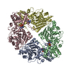
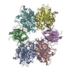
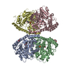
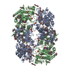
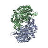
- PDB-1ecc: ESCHERICHIA COLI GLUTAMINE PHOSPHORIBOSYLPYROPHOSPHATE (PRPP) AMI... -
+ Open data

- Basic information
| Entry | Database: PDB / ID: 1ecc | ||||||
|---|---|---|---|---|---|---|---|
| Title | ESCHERICHIA COLI GLUTAMINE PHOSPHORIBOSYLPYROPHOSPHATE (PRPP) AMIDOTRANSFERASE COMPLEXED WITH MN-CPRPP AND 5-OXO-NORLEUCINE | ||||||
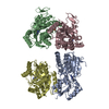
 Components Components | GLUTAMINE PHOSPHORIBOSYLPYROPHOSPHATE AMIDOTRANSFERASE | ||||||
 Keywords Keywords |  TRANSFERASE / GLUTAMINE AMIDOTRANSFERASE / PURINE BIOSYNTHESIS / GLYCOSYLTRANSFERASE TRANSFERASE / GLUTAMINE AMIDOTRANSFERASE / PURINE BIOSYNTHESIS / GLYCOSYLTRANSFERASE | ||||||
| Function / homology |  Function and homology informationamidophosphoribosyltransferase / amidophosphoribosyltransferase activity / purine nucleobase biosynthetic process / purine nucleotide biosynthetic process / 'de novo' IMP biosynthetic process / guanosine tetraphosphate binding / glutamine metabolic process / glycosyltransferase activity / magnesium ion binding / identical protein binding ...amidophosphoribosyltransferase / amidophosphoribosyltransferase activity / purine nucleobase biosynthetic process / purine nucleotide biosynthetic process / 'de novo' IMP biosynthetic process / guanosine tetraphosphate binding / glutamine metabolic process / glycosyltransferase activity / magnesium ion binding / identical protein binding / cytosol / cytoplasm Function and homology informationamidophosphoribosyltransferase / amidophosphoribosyltransferase activity / purine nucleobase biosynthetic process / purine nucleotide biosynthetic process / 'de novo' IMP biosynthetic process / guanosine tetraphosphate binding / glutamine metabolic process / glycosyltransferase activity / magnesium ion binding / identical protein binding ...amidophosphoribosyltransferase / amidophosphoribosyltransferase activity / purine nucleobase biosynthetic process / purine nucleotide biosynthetic process / 'de novo' IMP biosynthetic process / guanosine tetraphosphate binding / glutamine metabolic process / glycosyltransferase activity / magnesium ion binding / identical protein binding / cytosol / cytoplasmSimilarity search - Function | ||||||
| Biological species |  Escherichia coli (E. coli) Escherichia coli (E. coli) | ||||||
| Method | X-RAY DIFFRACTION / MOLECULAR REPLACEMENT / Resolution: 2.4 Å | ||||||
 Authors Authors | Krahn, J.M. / Smith, J.L. | ||||||
 Citation Citation | Journal: Biochemistry / Year: 1997 Title: Coupled formation of an amidotransferase interdomain ammonia channel and a phosphoribosyltransferase active site. Authors: Krahn, J.M. / Kim, J.H. / Burns, M.R. / Parry, R.J. / Zalkin, H. / Smith, J.L. | ||||||
| History |
|
- Structure visualization
| Structure viewer | Molecule: MolmilJmol/JSmol |
|---|
- Downloads & links
-Download
| PDBx/mmCIF format | 1ecc.cif.gz | 206.8 KB | Display | PDBx/mmCIF format |
|---|---|---|---|---|
| PDB format | pdb1ecc.ent.gz | 168.3 KB | Display | PDB format |
| PDBx/mmJSON format | 1ecc.json.gz | Tree view | PDBx/mmJSON format | |
| Others |  Other downloads Other downloads |
-Validation report
| Arichive directory | https://data.pdbj.org/pub/pdb/validation_reports/ec/1eccftp://data.pdbj.org/pub/pdb/validation_reports/ec/1ecc | HTTPS FTP |
|---|
-Related structure data
| Related structure data |  1ecbC 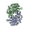 1ecfS S: Starting model for refinement C: citing same article ( |
|---|---|
| Similar structure data |
-Links
 PDBj
PDBj


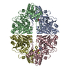
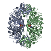
- Assembly
| Deposited unit | 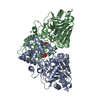
| ||||||||
|---|---|---|---|---|---|---|---|---|---|
| 1 | 
| ||||||||
| Unit cell |
| ||||||||
| Noncrystallographic symmetry (NCS) | NCS oper: (Code: given Matrix: (-0.142832, 0.84025, 0.523047), Vector : |
-Components
| #1: Protein | Mass: 56422.684 Da / Num. of mol.: 2 Source method: isolated from a genetically manipulated source Source: (gene. exp.) Escherichia coli (E. coli) / Gene: PURF / Plasmid: PT7F1 / Species (production host): Escherichia coli / Production host: Escherichia coli BL21(DE3) (bacteria) / Strain (production host): BL21 (DE3)References: UniProt: P00496, UniProt: P0AG16*PLUS, amidophosphoribosyltransferase#2: Chemical | ChemComp-MN /   Mass: 54.938 Da / Num. of mol.: 5 / Source method: obtained synthetically / Formula: Mn Mass: 54.938 Da / Num. of mol.: 5 / Source method: obtained synthetically / Formula: Mn#3: Chemical | 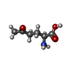  Type: L-peptide linking / Mass: 145.156 Da / Num. of mol.: 2 / Source method: obtained synthetically / Formula: C6H11NO3 Type: L-peptide linking / Mass: 145.156 Da / Num. of mol.: 2 / Source method: obtained synthetically / Formula: C6H11NO3#4: Chemical | 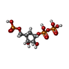  Mass: 388.097 Da / Num. of mol.: 2 / Source method: obtained synthetically / Formula: C6H15O13P3 Mass: 388.097 Da / Num. of mol.: 2 / Source method: obtained synthetically / Formula: C6H15O13P3#5: Water | ChemComp-HOH / | Water Mass: 18.015 Da / Num. of mol.: 291 / Source method: isolated from a natural source / Formula: H2O Mass: 18.015 Da / Num. of mol.: 291 / Source method: isolated from a natural source / Formula: H2ONonpolymer details | A WELL ORDERED MN++ EXISTS AT A LATTICE CONTACT. THIS METAL IS LIGATED TO GLU B 449, ASP B 471, AND ...A WELL ORDERED MN++ EXISTS AT A LATTICE CONTACT. THIS METAL IS LIGATED TO GLU B 449, ASP B 471, AND WATERS HOH A 507, HOH A 508, HOH B 507, AND HOH B 508. LIGATION TO ASP B 471 IS TO A SYMMETRY-EQUIVALENT | |
|---|
-Experimental details
-Experiment
| Experiment | Method: X-RAY DIFFRACTION / Number of used crystals: 1 |
|---|
- Sample preparation
| Crystal | Density Matthews: 2.29 Å3/Da / Density % sol: 46 % Description: A SINGLE GAT DOMAIN (249 RESIDUES) WAS USED AS THE PROBE MODEL DUE TO ALLOSTERIC DIFFERENCES BETWEEN THIS STRUCTURE AND THAT OF 1ECF. | ||||||||||||||||||||||||||||||||||||||||||||||||||||||||||||
|---|---|---|---|---|---|---|---|---|---|---|---|---|---|---|---|---|---|---|---|---|---|---|---|---|---|---|---|---|---|---|---|---|---|---|---|---|---|---|---|---|---|---|---|---|---|---|---|---|---|---|---|---|---|---|---|---|---|---|---|---|---|
| Crystal grow | pH: 5.8 Details: 0.42MM CPRPP, 25MM MNCL2, 40MM BIS-TRIS PH 5.8, 3.2% 2-PROPANOL, 7.5% PEG-3350 | ||||||||||||||||||||||||||||||||||||||||||||||||||||||||||||
| Crystal | *PLUS | ||||||||||||||||||||||||||||||||||||||||||||||||||||||||||||
| Crystal grow | *PLUS Temperature: 20 ℃ / Method: vapor diffusion | ||||||||||||||||||||||||||||||||||||||||||||||||||||||||||||
| Components of the solutions | *PLUS
|
-Data collection
| Diffraction | Mean temperature: 110 K |
|---|---|
| Diffraction source | Source: ROTATING ANODE / Type: RIGAKU RUH2R / Wavelength: 1.5418 |
| Detector | Type: RIGAKU RAXIS IIC / Detector: IMAGE PLATE / Date: Aug 15, 1996 |
| Radiation | Monochromator: MSC DOUBLE MIRROR / Monochromatic (M) / Laue (L): M / Scattering type: x-ray |
| Radiation wavelength | Wavelength: 1.5418 Å / Relative weight: 1 |
| Reflection | Resolution: 2.4→25 Å / Num. obs: 27331 / % possible obs: 88.3 % / Observed criterion σ(I): -3 / Redundancy: 2.8 % / Biso Wilson estimate: 42.9 Å2 / Rsym value: 0.036 / Net I/σ(I): 22.7 |
| Reflection shell | Resolution: 2.4→2.5 Å / Mean I/σ(I) obs: 3.1 / Rsym value: 0.172 / % possible all: 60.3 |
| Reflection | *PLUS Num. measured all: 99285 / Rmerge(I) obs: 0.036 |
| Reflection shell | *PLUS % possible obs: 60.3 % / Rmerge(I) obs: 0.172 |
- Processing
| Software |
| ||||||||||||||||||||||||||||||||||||||||||||||||||||||||||||||||||||||||||||||||
|---|---|---|---|---|---|---|---|---|---|---|---|---|---|---|---|---|---|---|---|---|---|---|---|---|---|---|---|---|---|---|---|---|---|---|---|---|---|---|---|---|---|---|---|---|---|---|---|---|---|---|---|---|---|---|---|---|---|---|---|---|---|---|---|---|---|---|---|---|---|---|---|---|---|---|---|---|---|---|---|---|---|
| Refinement | Method to determine structure: MOLECULAR REPLACEMENT Starting model: PDB ENTRY 1ECF Resolution: 2.4→25 Å / Data cutoff high absF: 1000000 / Data cutoff low absF: 0 / Cross valid method: FREE R / σ(F): 0 Details: ADDITIONAL TOPOLOGY/PARAMETER FILES FOR THE CYS/ONL LINKAGE WERE USED, WHICH HAVE A NOMENCLATURE DIFFERENT FROM THE ONL RESIDUE IN THIS ENTRY.
| ||||||||||||||||||||||||||||||||||||||||||||||||||||||||||||||||||||||||||||||||
| Displacement parameters | Biso mean: 34.3 Å2
| ||||||||||||||||||||||||||||||||||||||||||||||||||||||||||||||||||||||||||||||||
| Refine analyze | Luzzati coordinate error obs: 0.27 Å / Luzzati d res low obs: 5 Å | ||||||||||||||||||||||||||||||||||||||||||||||||||||||||||||||||||||||||||||||||
| Refinement step | Cycle: LAST / Resolution: 2.4→25 Å
| ||||||||||||||||||||||||||||||||||||||||||||||||||||||||||||||||||||||||||||||||
| Refine LS restraints |
| ||||||||||||||||||||||||||||||||||||||||||||||||||||||||||||||||||||||||||||||||
| LS refinement shell | Resolution: 2.4→2.49 Å / Total num. of bins used: 10
| ||||||||||||||||||||||||||||||||||||||||||||||||||||||||||||||||||||||||||||||||
| Xplor file |
| ||||||||||||||||||||||||||||||||||||||||||||||||||||||||||||||||||||||||||||||||
| Software | *PLUS Name: X-PLOR / Version: 3.8 / Classification: refinement | ||||||||||||||||||||||||||||||||||||||||||||||||||||||||||||||||||||||||||||||||
| Refine LS restraints | *PLUS
| ||||||||||||||||||||||||||||||||||||||||||||||||||||||||||||||||||||||||||||||||
| LS refinement shell | *PLUS Rfactor Rfree: 0.35 |
