 Movie
Movie Controller

[English] 日本語
 Yorodumi
- PDB-1bi8: MECHANISM OF G1 CYCLIN DEPENDENT KINASE INHIBITION FROM THE STRUC... -
+ Open data

- Basic information
| Entry | Database: PDB / ID: 1bi8 | ||||||
|---|---|---|---|---|---|---|---|
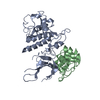
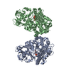
| Title | MECHANISM OF G1 CYCLIN DEPENDENT KINASE INHIBITION FROM THE STRUCTURES CDK6-P19INK4D INHIBITOR COMPLEX | ||||||
 Components Components |
| ||||||
 Keywords Keywords | COMPLEX (KINASE/INHIBITOR) /  CYCLIN DEPENDENT KINASE / CYCLIN DEPENDENT KINASE INHIBITORY PROTEIN / CDK / INK4 / CELL CYCLE / COMPLEX (KINASE-INHIBITOR) / COMPLEX (KINASE-INHIBITOR) complex CYCLIN DEPENDENT KINASE / CYCLIN DEPENDENT KINASE INHIBITORY PROTEIN / CDK / INK4 / CELL CYCLE / COMPLEX (KINASE-INHIBITOR) / COMPLEX (KINASE-INHIBITOR) complex | ||||||
| Function / homology |  Function and homology information Function and homology informationcyclin D2-CDK6 complex / cyclin D2-CDK4 complex / cell dedifferentiation / Drug-mediated inhibition of CDK4/CDK6 activity / Evasion of Oncogene Induced Senescence Due to Defective p16INK4A binding to CDK4 and CDK6 / Evasion of Oxidative Stress Induced Senescence Due to Defective p16INK4A binding to CDK4 and CDK6 / FBXO family protein binding / autophagic cell death / lateral ventricle development / negative regulation of cell cycle G1/S phase transition ...cyclin D2-CDK6 complex / cyclin D2-CDK4 complex / cell dedifferentiation / Drug-mediated inhibition of CDK4/CDK6 activity / Evasion of Oncogene Induced Senescence Due to Defective p16INK4A binding to CDK4 and CDK6 / Evasion of Oxidative Stress Induced Senescence Due to Defective p16INK4A binding to CDK4 and CDK6 / FBXO family protein binding / autophagic cell death / lateral ventricle development / negative regulation of cell cycle G1/S phase transition / negative regulation of myeloid cell differentiation / type B pancreatic cell development / negative regulation of monocyte differentiation / astrocyte development / dentate gyrus development / negative regulation of phosphorylation / negative regulation of intrinsic apoptotic signaling pathway in response to DNA damage / cyclin-dependent protein serine/threonine kinase inhibitor activity / response to vitamin D / gliogenesis / regulation of cell motility / Regulation of RUNX1 Expression and Activity / regulation of hematopoietic stem cell differentiation / positive regulation of cell-matrix adhesion / generation of neurons / DNA synthesis involved in DNA repair / regulation of cyclin-dependent protein serine/threonine kinase activity / regulation of G1/S transition of mitotic cell cycle / Defective binding of RB1 mutants to E2F1,(E2F2, E2F3) / negative regulation of cellular senescence / negative regulation of cell differentiation / negative regulation of cell cycle / hematopoietic stem cell differentiation / cyclin-dependent protein kinase holoenzyme complex / negative regulation of osteoblast differentiation / cyclin-dependent kinase / cyclin-dependent protein serine/threonine kinase activity / response to UV / response to retinoic acid / Notch signaling pathway / ruffle / regulation of G2/M transition of mitotic cell cycle / cyclin binding / : / sensory perception of sound / response to virus / negative regulation of cysteine-type endopeptidase activity involved in apoptotic process / regulation of erythrocyte differentiation / Oncogene Induced Senescence / G1/S transition of mitotic cell cycle / negative regulation of cell growth / Cyclin D associated events in G1 / negative regulation of epithelial cell proliferation / positive regulation of fibroblast proliferation / T cell differentiation in thymus / Senescence-Associated Secretory Phenotype (SASP) / regulation of gene expression / Oxidative Stress Induced Senescence / regulation of cell cycle / cell cycle / negative regulation of cell population proliferation / cell division / protein phosphorylation / protein serine kinase activity / centrosome / positive regulation of gene expression / protein kinase binding / negative regulation of transcription by RNA polymerase II / signal transduction / nucleoplasm / ATP binding / nucleus / cytosol / cytoplasmSimilarity search - Function | ||||||
| Biological species |  Homo sapiens (human) Homo sapiens (human) | ||||||
| Method | X-RAY DIFFRACTION / SYNCHROTRON / MOLECULAR REPLACEMENT / Resolution: 2.8 Å | ||||||
 Authors Authors | Russo, A.A. / Tong, L. / Lee, J.O. / Jeffrey, P.D. / Pavletich, N.P. | ||||||
 Citation Citation | Journal: Nature / Year: 1998 Title: Structural basis for inhibition of the cyclin-dependent kinase Cdk6 by the tumour suppressor p16INK4a. Authors: Russo, A.A. / Tong, L. / Lee, J.O. / Jeffrey, P.D. / Pavletich, N.P. | ||||||
| History |
|
- Structure visualization
| Structure viewer | Molecule: MolmilJmol/JSmol |
|---|
- Downloads & links
-Download
| PDBx/mmCIF format | 1bi8.cif.gz | 169.6 KB | Display | PDBx/mmCIF format |
|---|---|---|---|---|
| PDB format | pdb1bi8.ent.gz | 135.9 KB | Display | PDB format |
| PDBx/mmJSON format | 1bi8.json.gz | Tree view | PDBx/mmJSON format | |
| Others |  Other downloads Other downloads |
-Validation report
| Arichive directory | https://data.pdbj.org/pub/pdb/validation_reports/bi/1bi8ftp://data.pdbj.org/pub/pdb/validation_reports/bi/1bi8 | HTTPS FTP |
|---|
-Related structure data


-Links
 PDBj
PDBj


- Assembly
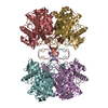
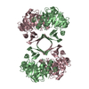
| Deposited unit | 
| ||||||||
|---|---|---|---|---|---|---|---|---|---|
| 1 |
| ||||||||
| Unit cell |
| ||||||||
| Noncrystallographic symmetry (NCS) | NCS oper: (Code: given Matrix: (-0.965395, 0.252602, 0.064847), Vector : |
-Components
| #1: Protein | / CDK6 Mass: 36987.328 Da / Num. of mol.: 2 Source method: isolated from a genetically manipulated source Source: (gene. exp.) Homo sapiens (human) / Cell line: SPODOPTERA FRUGIPERDA / Production host:   Spodoptera frugiperda (fall armyworm) / Strain (production host): HI5 Spodoptera frugiperda (fall armyworm) / Strain (production host): HI5References: UniProt: Q00534, Transferases; Transferring phosphorus-containing groups; Phosphotransferases with an alcohol group as acceptor#2: Protein | Cyclin-dependent kinase inhibitor protein / P19INK4DMass: 17723.166 Da / Num. of mol.: 2 Source method: isolated from a genetically manipulated source Source: (gene. exp.) Homo sapiens (human) / Cell line: SPODOPTERA FRUGIPERDA / Production host:  Escherichia coli (E. coli) / Strain (production host): HI5 / References: UniProt: P55273 Escherichia coli (E. coli) / Strain (production host): HI5 / References: UniProt: P55273 |
|---|
-Experimental details
-Experiment
| Experiment | Method: X-RAY DIFFRACTION / Number of used crystals: 1 |
|---|
- Sample preparation
| Crystal | Density Matthews: 3.16 Å3/Da / Density % sol: 61.07 % | |||||||||||||||||||||||||
|---|---|---|---|---|---|---|---|---|---|---|---|---|---|---|---|---|---|---|---|---|---|---|---|---|---|---|
| Crystal grow | *PLUS Temperature: 4 ℃ / pH: 6.5 / Method: vapor diffusion, hanging drop | |||||||||||||||||||||||||
| Components of the solutions | *PLUS
|
-Data collection
| Diffraction | Mean temperature: 110 K |
|---|---|
| Diffraction source | Source: SYNCHROTRON / Site: NSLS  / Beamline: X4A / Wavelength: 1.006 / Beamline: X4A / Wavelength: 1.006 |
| Detector | Type: RIGAKU / Detector: IMAGE PLATE / Date: Jan 1, 1998 / Details: MIRRORS |
| Radiation | Monochromator: MIRRORS / Monochromatic (M) / Laue (L): M / Scattering type: x-ray |
| Radiation wavelength | Wavelength: 1.006 Å / Relative weight: 1 |
| Reflection | Resolution: 2.8→20 Å / Num. obs: 33400 / % possible obs: 96.9 % / Observed criterion σ(I): 0 / Redundancy: 2.65 % / Rsym value: 0.055 / Net I/σ(I): 14.5 |
| Reflection shell | Resolution: 2.8→2.9 Å / Redundancy: 2.3 % / Mean I/σ(I) obs: 4.1 / Rsym value: 0.32 / % possible all: 91.3 |
| Reflection | *PLUS Num. measured all: 88741 / Rmerge(I) obs: 0.055 |
| Reflection shell | *PLUS % possible obs: 91.3 % / Rmerge(I) obs: 0.32 |
- Processing
| Software |
| ||||||||||||||||||||||||||||||||||||||||||||||||||||||||||||||||||||||||||||||||
|---|---|---|---|---|---|---|---|---|---|---|---|---|---|---|---|---|---|---|---|---|---|---|---|---|---|---|---|---|---|---|---|---|---|---|---|---|---|---|---|---|---|---|---|---|---|---|---|---|---|---|---|---|---|---|---|---|---|---|---|---|---|---|---|---|---|---|---|---|---|---|---|---|---|---|---|---|---|---|---|---|---|
| Refinement | Method to determine structure: MOLECULAR REPLACEMENT Starting model: BNL-22910 Resolution: 2.8→10 Å / Isotropic thermal model: RESTRAINED / Cross valid method: THROUGHOUT / σ(F): 1 / Details: REFINED WITH NCS STRICT THROUGHOUT
| ||||||||||||||||||||||||||||||||||||||||||||||||||||||||||||||||||||||||||||||||
| Displacement parameters |
| ||||||||||||||||||||||||||||||||||||||||||||||||||||||||||||||||||||||||||||||||
| Refinement step | Cycle: LAST / Resolution: 2.8→10 Å
| ||||||||||||||||||||||||||||||||||||||||||||||||||||||||||||||||||||||||||||||||
| Refine LS restraints |
| ||||||||||||||||||||||||||||||||||||||||||||||||||||||||||||||||||||||||||||||||
| Refine LS restraints NCS | NCS model details: STRICT | ||||||||||||||||||||||||||||||||||||||||||||||||||||||||||||||||||||||||||||||||
| Software | *PLUS Name: X-PLOR / Version: 3.8 / Classification: refinement | ||||||||||||||||||||||||||||||||||||||||||||||||||||||||||||||||||||||||||||||||
| Refinement | *PLUS | ||||||||||||||||||||||||||||||||||||||||||||||||||||||||||||||||||||||||||||||||
| Solvent computation | *PLUS | ||||||||||||||||||||||||||||||||||||||||||||||||||||||||||||||||||||||||||||||||
| Displacement parameters | *PLUS |
