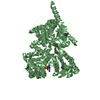
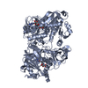
 Movie
Movie Controller

[English] 日本語
 Yorodumi
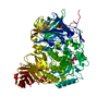
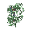
- PDB-1bi7: MECHANISM OF G1 CYCLIN DEPENDENT KINASE INHIBITION FROM THE STRUC... -
+ Open data

- Basic information
| Entry | Database: PDB / ID: 1bi7 | ||||||
|---|---|---|---|---|---|---|---|
| Title | MECHANISM OF G1 CYCLIN DEPENDENT KINASE INHIBITION FROM THE STRUCTURE OF THE CDK6-P16INK4A TUMOR SUPPRESSOR COMPLEX | ||||||
 Components Components |
| ||||||
 Keywords Keywords | COMPLEX (KINASE/ANTI-ONCOGENE) /  CYCLIN DEPENDENT KINASE / CYCLIN DEPENDENT KINASE INHIBITORY PROTEIN / CDK / INK4 / CELL CYCLE / MULTIPLE TUMOR SUPPRESSOR / MTS1 / COMPLEX (KINASE-ANTI-ONCOGENE) / COMPLEX (KINASE-ANTI-ONCOGENE) complex CYCLIN DEPENDENT KINASE / CYCLIN DEPENDENT KINASE INHIBITORY PROTEIN / CDK / INK4 / CELL CYCLE / MULTIPLE TUMOR SUPPRESSOR / MTS1 / COMPLEX (KINASE-ANTI-ONCOGENE) / COMPLEX (KINASE-ANTI-ONCOGENE) complex | ||||||
| Function / homology |  Function and homology information Function and homology informationsenescence-associated heterochromatin focus / positive regulation of macrophage apoptotic process / cyclin D2-CDK6 complex / Evasion of Oncogene Induced Senescence Due to Defective p16INK4A binding to CDK4 / Evasion of Oxidative Stress Induced Senescence Due to Defective p16INK4A binding to CDK4 / cell dedifferentiation / Drug-mediated inhibition of CDK4/CDK6 activity / Evasion of Oncogene Induced Senescence Due to Defective p16INK4A binding to CDK4 and CDK6 / Evasion of Oxidative Stress Induced Senescence Due to Defective p16INK4A binding to CDK4 and CDK6 / FBXO family protein binding ...senescence-associated heterochromatin focus / positive regulation of macrophage apoptotic process / cyclin D2-CDK6 complex / Evasion of Oncogene Induced Senescence Due to Defective p16INK4A binding to CDK4 / Evasion of Oxidative Stress Induced Senescence Due to Defective p16INK4A binding to CDK4 / cell dedifferentiation / Drug-mediated inhibition of CDK4/CDK6 activity / Evasion of Oncogene Induced Senescence Due to Defective p16INK4A binding to CDK4 and CDK6 / Evasion of Oxidative Stress Induced Senescence Due to Defective p16INK4A binding to CDK4 and CDK6 / FBXO family protein binding / lateral ventricle development / negative regulation of myeloid cell differentiation / positive regulation of smooth muscle cell apoptotic process / type B pancreatic cell development / negative regulation of monocyte differentiation / astrocyte development / negative regulation of phosphorylation / dentate gyrus development / negative regulation of cyclin-dependent protein serine/threonine kinase activity / gliogenesis / cyclin-dependent protein serine/threonine kinase inhibitor activity / regulation of cell motility / Regulation of RUNX1 Expression and Activity / regulation of hematopoietic stem cell differentiation / positive regulation of cell-matrix adhesion / generation of neurons / negative regulation of cell-matrix adhesion / negative regulation of NF-kappaB transcription factor activity / regulation of G1/S transition of mitotic cell cycle / Defective binding of RB1 mutants to E2F1,(E2F2, E2F3) / negative regulation of cellular senescence / negative regulation of cell differentiation / Transcriptional Regulation by VENTX / negative regulation of cell cycle / NF-kappaB binding / replicative senescence / cyclin-dependent protein kinase holoenzyme complex / hematopoietic stem cell differentiation / negative regulation of osteoblast differentiation / cyclin-dependent kinase / cyclin-dependent protein serine/threonine kinase activity / Notch signaling pathway / ruffle / regulation of G2/M transition of mitotic cell cycle / cyclin binding / response to organic substance / G1/S transition of mitotic cell cycle / response to virus / regulation of erythrocyte differentiation / Oncogene Induced Senescence / negative regulation of cell growth / Cyclin D associated events in G1 / positive regulation of fibroblast proliferation / negative regulation of epithelial cell proliferation / cellular senescence / T cell differentiation in thymus / Senescence-Associated Secretory Phenotype (SASP) / regulation of gene expression / Oxidative Stress Induced Senescence / Ras protein signal transduction / regulation of cell cycle / cell cycle / cell division / negative regulation of cell population proliferation / protein phosphorylation / protein serine kinase activity / negative regulation of DNA-templated transcription / centrosome / positive regulation of gene expression / protein kinase binding / negative regulation of transcription by RNA polymerase II / signal transduction / RNA binding / nucleoplasm / ATP binding / nucleus / cytosol / cytoplasmSimilarity search - Function | ||||||
| Biological species |  Homo sapiens (human) Homo sapiens (human) | ||||||
| Method | X-RAY DIFFRACTION / SYNCHROTRON / MIR, MAD / Resolution: 3.4 Å | ||||||
 Authors Authors | Russo, A.A. / Tong, L. / Lee, J.O. / Jeffrey, P.D. / Pavletich, N.P. | ||||||
 Citation Citation | Journal: Nature / Year: 1998 Title: Structural basis for inhibition of the cyclin-dependent kinase Cdk6 by the tumour suppressor p16INK4a. Authors: Russo, A.A. / Tong, L. / Lee, J.O. / Jeffrey, P.D. / Pavletich, N.P. | ||||||
| History |
|
- Structure visualization
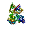
| Structure viewer | Molecule: MolmilJmol/JSmol |
|---|
- Downloads & links
-Download
| PDBx/mmCIF format | 1bi7.cif.gz | 81.6 KB | Display | PDBx/mmCIF format |
|---|---|---|---|---|
| PDB format | pdb1bi7.ent.gz | 62.3 KB | Display | PDB format |
| PDBx/mmJSON format | 1bi7.json.gz | Tree view | PDBx/mmJSON format | |
| Others |  Other downloads Other downloads |
-Validation report
| Arichive directory | https://data.pdbj.org/pub/pdb/validation_reports/bi/1bi7ftp://data.pdbj.org/pub/pdb/validation_reports/bi/1bi7 | HTTPS FTP |
|---|
-Related structure data


-Links
 PDBj
PDBj


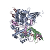
- Assembly
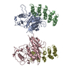
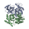
| Deposited unit | 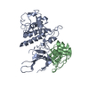
| ||||||||
|---|---|---|---|---|---|---|---|---|---|
| 1 | 
| ||||||||
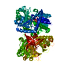
| Unit cell |
|
-Components
| #1: Protein | / CDK6 Mass: 36987.328 Da / Num. of mol.: 1 Source method: isolated from a genetically manipulated source Source: (gene. exp.) Homo sapiens (human) / Cell line: SPODOPTERA FRUGIPERDA / Production host:   Spodoptera frugiperda (fall armyworm) / Strain (production host): HI5 Spodoptera frugiperda (fall armyworm) / Strain (production host): HI5References: UniProt: Q00534, Transferases; Transferring phosphorus-containing groups; Phosphotransferases with an alcohol group as acceptor |
|---|---|
| #2: Protein | Mass: 16555.586 Da / Num. of mol.: 1 Source method: isolated from a genetically manipulated source Source: (gene. exp.) Homo sapiens (human) / Cell line: SPODOPTERA FRUGIPERDA / Production host:  Escherichia coli (E. coli) / Strain (production host): HI5 / References: UniProt: P42771 Escherichia coli (E. coli) / Strain (production host): HI5 / References: UniProt: P42771 |
-Experimental details
-Experiment
| Experiment | Method: X-RAY DIFFRACTION / Number of used crystals: 2 |
|---|
- Sample preparation
| Crystal | Density Matthews: 3.98 Å3/Da / Density % sol: 69.08 % | ||||||||||||||||||||
|---|---|---|---|---|---|---|---|---|---|---|---|---|---|---|---|---|---|---|---|---|---|
| Crystal grow | *PLUS Temperature: 4 ℃ / pH: 7.5 / Method: vapor diffusion, hanging drop | ||||||||||||||||||||
| Components of the solutions | *PLUS
|
-Data collection
| Diffraction | Mean temperature: 110 K |
|---|---|
| Diffraction source | Source: SYNCHROTRON / Site: CHESS  / Beamline: A1 / Wavelength: 0.908 / Beamline: A1 / Wavelength: 0.908 |
| Detector | Detector: CCD / Date: Jan 1, 1997 / Details: MIRRORS |
| Radiation | Monochromator: MIRRORS / Monochromatic (M) / Laue (L): M / Scattering type: x-ray |
| Radiation wavelength | Wavelength: 0.908 Å / Relative weight: 1 |
| Reflection | Resolution: 3.4→20 Å / Num. obs: 12443 / % possible obs: 97.7 % / Observed criterion σ(I): 0 / Redundancy: 5.5 % / Rsym value: 0.068 / Net I/σ(I): 17.7 |
| Reflection shell | Resolution: 3.4→3.52 Å / Redundancy: 4.1 % / Mean I/σ(I) obs: 6.9 / Rsym value: 0.33 / % possible all: 96.2 |
| Reflection | *PLUS Num. measured all: 68633 / Rmerge(I) obs: 0.068 |
| Reflection shell | *PLUS % possible obs: 96.2 % / Rmerge(I) obs: 0.33 |
- Processing
| Software |
| ||||||||||||||||||||||||||||||||||||||||||||||||||||||||||||
|---|---|---|---|---|---|---|---|---|---|---|---|---|---|---|---|---|---|---|---|---|---|---|---|---|---|---|---|---|---|---|---|---|---|---|---|---|---|---|---|---|---|---|---|---|---|---|---|---|---|---|---|---|---|---|---|---|---|---|---|---|---|
| Refinement | Method to determine structure: MIR, MAD / Resolution: 3.4→10 Å / Isotropic thermal model: GROUP / Cross valid method: THROUGHOUT / σ(F): 1
| ||||||||||||||||||||||||||||||||||||||||||||||||||||||||||||
| Refinement step | Cycle: LAST / Resolution: 3.4→10 Å
| ||||||||||||||||||||||||||||||||||||||||||||||||||||||||||||
| Refine LS restraints |
| ||||||||||||||||||||||||||||||||||||||||||||||||||||||||||||
| Software | *PLUS Name: X-PLOR / Version: 3.8 / Classification: refinement | ||||||||||||||||||||||||||||||||||||||||||||||||||||||||||||
| Refinement | *PLUS | ||||||||||||||||||||||||||||||||||||||||||||||||||||||||||||
| Solvent computation | *PLUS | ||||||||||||||||||||||||||||||||||||||||||||||||||||||||||||
| Displacement parameters | *PLUS |
