 Movie
Movie Controller


+ Open data

- Basic information
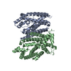
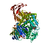
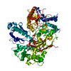
| Entry | Database: PDB / ID: 4rie | ||||||
|---|---|---|---|---|---|---|---|
| Title | Landomycin Glycosyltransferase LanGT2 | ||||||
 Components Components | Glycosyl transferase homolog | ||||||
 Keywords Keywords |  TRANSFERASE / GT fold / glycosyltransferase TRANSFERASE / GT fold / glycosyltransferase | ||||||
| Function / homology |  Function and homology information Function and homology informationUDP-glycosyltransferase activity / hexosyltransferase activity / antibiotic biosynthetic processSimilarity search - Function | ||||||
| Biological species |  Streptomyces cyanogenus (bacteria) Streptomyces cyanogenus (bacteria) | ||||||
| Method | X-RAY DIFFRACTION / SYNCHROTRON / MOLECULAR REPLACEMENT / Resolution: 2.162 Å | ||||||
 Authors Authors | Tam, H.K. / Gerhardt, S. / Breit, B. / Bechthold, A. / Einsle, O. | ||||||
 Citation Citation | Journal: Angew.Chem.Int.Ed.Engl. / Year: 2015 Title: Structural Characterization of O- and C-Glycosylating Variants of the Landomycin Glycosyltransferase LanGT2. Authors: Tam, H.K. / Harle, J. / Gerhardt, S. / Rohr, J. / Wang, G. / Thorson, J.S. / Bigot, A. / Lutterbeck, M. / Seiche, W. / Breit, B. / Bechthold, A. / Einsle, O. | ||||||
| History |
|
- Structure visualization
| Structure viewer | Molecule: MolmilJmol/JSmol |
|---|
- Downloads & links
-Download
| PDBx/mmCIF format | 4rie.cif.gz | 272.5 KB | Display | PDBx/mmCIF format |
|---|---|---|---|---|
| PDB format | pdb4rie.ent.gz | 221.9 KB | Display | PDB format |
| PDBx/mmJSON format | 4rie.json.gz | Tree view | PDBx/mmJSON format | |
| Others |  Other downloads Other downloads |
-Validation report
| Arichive directory | https://data.pdbj.org/pub/pdb/validation_reports/ri/4rieftp://data.pdbj.org/pub/pdb/validation_reports/ri/4rie | HTTPS FTP |
|---|
-Related structure data
| Related structure data | 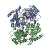 4rifC 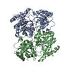 4rigC 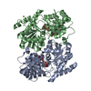 4rihC 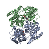 4riiC  2p6pS S: Starting model for refinement C: citing same article ( |
|---|---|
| Similar structure data |


-Links
 PDBj
PDBj- Assembly
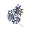
| Deposited unit | 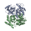
| ||||||||||||
|---|---|---|---|---|---|---|---|---|---|---|---|---|---|
| 1 |
| ||||||||||||
| Unit cell |
| ||||||||||||
| Noncrystallographic symmetry (NCS) | NCS oper:
|
-Components
| #1: Protein | Mass: 40406.105 Da / Num. of mol.: 2 / Fragment: Glycosyltransferase Source method: isolated from a genetically manipulated source Source: (gene. exp.) Streptomyces cyanogenus (bacteria) / Strain: S136 / Gene: lanGT2 / Plasmid: pET21::langt2 / Production host: Escherichia coli (E. coli) / Strain (production host): BL21(DE3)pLysS / References: UniProt: Q9ZGC0#2: Water | ChemComp-HOH / | Water Mass: 18.015 Da / Num. of mol.: 131 / Source method: isolated from a natural source / Formula: H2O Mass: 18.015 Da / Num. of mol.: 131 / Source method: isolated from a natural source / Formula: H2O |
|---|
-Experimental details
-Experiment
| Experiment | Method: X-RAY DIFFRACTION / Number of used crystals: 1 |
|---|
- Sample preparation
| Crystal | Density Matthews: 2.21 Å3/Da / Density % sol: 44.36 % |
|---|---|
| Crystal grow | Temperature: 293 K / Method: vapor diffusion, sitting drop / pH: 6.8 Details: 1.3 M sodium citrate 0.1 M HEPES/NaOH, pH 6.8, VAPOR DIFFUSION, SITTING DROP, temperature 293K |
-Data collection
| Diffraction | Mean temperature: 100 K |
|---|---|
| Diffraction source | Source: SYNCHROTRON / Site: SLS  / Beamline: X06SA / Wavelength: 1 Å / Beamline: X06SA / Wavelength: 1 Å |
| Detector | Type: DECTRIS PILATUS 6M / Detector: PIXEL / Date: Jun 27, 2013 |
| Radiation | Monochromator: graphite / Protocol: SINGLE WAVELENGTH / Monochromatic (M) / Laue (L): M / Scattering type: x-ray |
| Radiation wavelength | Wavelength: 1 Å / Relative weight: 1 |
| Reflection | Resolution: 2.16→214.31 Å / Num. all: 37364 / Num. obs: 37364 / % possible obs: 100 % / Observed criterion σ(F): 2 / Observed criterion σ(I): 2.5 / Rmerge(I) obs: 0.043 / Net I/σ(I): 40.4 |
| Reflection shell | Resolution: 2.16→2.28 Å / Rmerge(I) obs: 0.49 / Mean I/σ(I) obs: 5.3 / % possible all: 100 |
- Processing
| Software |
| ||||||||||||||||||||||||||||||||||||||||||||||||||||||||||||||||||||||||||||||||||||||||||||||||||||||||||||||||||||||||||||||||||||||||||||||||||||||||||||||||||||||||||||||||||||||
|---|---|---|---|---|---|---|---|---|---|---|---|---|---|---|---|---|---|---|---|---|---|---|---|---|---|---|---|---|---|---|---|---|---|---|---|---|---|---|---|---|---|---|---|---|---|---|---|---|---|---|---|---|---|---|---|---|---|---|---|---|---|---|---|---|---|---|---|---|---|---|---|---|---|---|---|---|---|---|---|---|---|---|---|---|---|---|---|---|---|---|---|---|---|---|---|---|---|---|---|---|---|---|---|---|---|---|---|---|---|---|---|---|---|---|---|---|---|---|---|---|---|---|---|---|---|---|---|---|---|---|---|---|---|---|---|---|---|---|---|---|---|---|---|---|---|---|---|---|---|---|---|---|---|---|---|---|---|---|---|---|---|---|---|---|---|---|---|---|---|---|---|---|---|---|---|---|---|---|---|---|---|---|---|
| Refinement | Method to determine structure: MOLECULAR REPLACEMENT Starting model: 2P6P Resolution: 2.162→65.81 Å / Cor.coef. Fo:Fc: 0.955 / Cor.coef. Fo:Fc free: 0.926 / SU B: 12.692 / SU ML: 0.16 / Isotropic thermal model: isotropic / Cross valid method: THROUGHOUT / ESU R: 0.272 / ESU R Free: 0.208 / Stereochemistry target values: MAXIMUM LIKELIHOOD / Details: HYDROGENS HAVE BEEN USED IF PRESENT IN THE INPUT
| ||||||||||||||||||||||||||||||||||||||||||||||||||||||||||||||||||||||||||||||||||||||||||||||||||||||||||||||||||||||||||||||||||||||||||||||||||||||||||||||||||||||||||||||||||||||
| Solvent computation | Ion probe radii: 0.8 Å / Shrinkage radii: 0.8 Å / VDW probe radii: 1.2 Å / Solvent model: MASK | ||||||||||||||||||||||||||||||||||||||||||||||||||||||||||||||||||||||||||||||||||||||||||||||||||||||||||||||||||||||||||||||||||||||||||||||||||||||||||||||||||||||||||||||||||||||
| Displacement parameters | Biso mean: 46.688 Å2
| ||||||||||||||||||||||||||||||||||||||||||||||||||||||||||||||||||||||||||||||||||||||||||||||||||||||||||||||||||||||||||||||||||||||||||||||||||||||||||||||||||||||||||||||||||||||
| Refinement step | Cycle: LAST / Resolution: 2.162→65.81 Å
| ||||||||||||||||||||||||||||||||||||||||||||||||||||||||||||||||||||||||||||||||||||||||||||||||||||||||||||||||||||||||||||||||||||||||||||||||||||||||||||||||||||||||||||||||||||||
| Refine LS restraints |
|