 Movie
Movie Controller


+ Open data

- Basic information
| Entry | Database: PDB / ID: 1b56 | ||||||
|---|---|---|---|---|---|---|---|
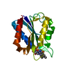
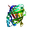
| Title | HUMAN RECOMBINANT EPIDERMAL FATTY ACID BINDING PROTEIN | ||||||
 Components Components | FATTY ACID BINDING PROTEIN Fatty acid-binding protein Fatty acid-binding protein | ||||||
 Keywords Keywords | LIPID BINDING PROTEIN / LIPID-BINDING / FATTY ACID TRANSPORT / BETA BARREL | ||||||
| Function / homology |  Function and homology information Function and homology informationregulation of prostaglandin biosynthetic process / regulation of retrograde trans-synaptic signaling by endocanabinoid / lipid transport across blood-brain barrier / positive regulation of peroxisome proliferator activated receptor signaling pathway / negative regulation of glucose transmembrane transport / regulation of sensory perception of pain / phosphatidylcholine biosynthetic process / retinoic acid binding / Signaling by Retinoic Acid / long-chain fatty acid transmembrane transporter activity ...regulation of prostaglandin biosynthetic process / regulation of retrograde trans-synaptic signaling by endocanabinoid / lipid transport across blood-brain barrier / positive regulation of peroxisome proliferator activated receptor signaling pathway / negative regulation of glucose transmembrane transport / regulation of sensory perception of pain / phosphatidylcholine biosynthetic process / retinoic acid binding / Signaling by Retinoic Acid / long-chain fatty acid transmembrane transporter activity / Triglyceride catabolism / epidermis development / fatty acid transport / long-chain fatty acid transport / secretory granule membrane / fatty acid binding / lipid metabolic process / glucose metabolic process / azurophil granule lumen / glucose homeostasis / positive regulation of cold-induced thermogenesis / postsynaptic density / synapse / lipid binding / Neutrophil degranulation / extracellular exosome / extracellular region / nucleoplasm / identical protein binding / nucleus / plasma membrane / cytosol / cytoplasmSimilarity search - Function | ||||||
| Biological species |  Homo sapiens (human) Homo sapiens (human) | ||||||
| Method | X-RAY DIFFRACTION / SYNCHROTRON / MOLECULAR REPLACEMENT / Resolution: 2.05 Å | ||||||
 Authors Authors | Van Tilbeurgh, H. / Hohoff, C. / Borchers, T. / Spener, F. | ||||||
 Citation Citation | Journal: Biochemistry / Year: 1999 Title: Expression, purification, and crystal structure determination of recombinant human epidermal-type fatty acid binding protein. Authors: Hohoff, C. / Borchers, T. / Rustow, B. / Spener, F. / van Tilbeurgh, H. | ||||||
| History |
|
- Structure visualization
| Structure viewer | Molecule: MolmilJmol/JSmol |
|---|
- Downloads & links
-Download
| PDBx/mmCIF format | 1b56.cif.gz | 41.3 KB | Display | PDBx/mmCIF format |
|---|---|---|---|---|
| PDB format | pdb1b56.ent.gz | 32.3 KB | Display | PDB format |
| PDBx/mmJSON format | 1b56.json.gz | Tree view | PDBx/mmJSON format | |
| Others |  Other downloads Other downloads |
-Validation report
| Arichive directory | https://data.pdbj.org/pub/pdb/validation_reports/b5/1b56ftp://data.pdbj.org/pub/pdb/validation_reports/b5/1b56 | HTTPS FTP |
|---|
-Related structure data
| Related structure data |  2hmbS S: Starting model for refinement |
|---|---|
| Similar structure data |


-Links
 PDBj
PDBj


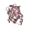
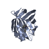
- Assembly
| Deposited unit | 
| ||||||||
|---|---|---|---|---|---|---|---|---|---|
| 1 |
| ||||||||
| Unit cell |
|
-Components
| #1: Protein | Fatty acid-binding protein Mass: 15185.457 Da / Num. of mol.: 1 Source method: isolated from a genetically manipulated source Source: (gene. exp.) Homo sapiens (human) / Organ: EPIDERMIS / Production host:  Escherichia coli (E. coli) / References: UniProt: Q01469 Escherichia coli (E. coli) / References: UniProt: Q01469 |
|---|---|
| #2: Chemical | ChemComp-PLM / Palmitic acid  Mass: 256.424 Da / Num. of mol.: 1 / Source method: obtained synthetically / Formula: C16H32O2 Mass: 256.424 Da / Num. of mol.: 1 / Source method: obtained synthetically / Formula: C16H32O2 |
| #3: Water | ChemComp-HOH / Water Mass: 18.015 Da / Num. of mol.: 39 / Source method: isolated from a natural source / Formula: H2O Mass: 18.015 Da / Num. of mol.: 39 / Source method: isolated from a natural source / Formula: H2O |
-Experimental details
-Experiment
| Experiment | Method: X-RAY DIFFRACTION / Number of used crystals: 1 |
|---|
- Sample preparation
| Crystal | Density Matthews: 2.55 Å3/Da / Density % sol: 52 % | ||||||||||||||||||||
|---|---|---|---|---|---|---|---|---|---|---|---|---|---|---|---|---|---|---|---|---|---|
| Crystal grow | pH: 6 / Details: pH 6.0 | ||||||||||||||||||||
| Crystal | *PLUS | ||||||||||||||||||||
| Crystal grow | *PLUS pH: 5.6 / Method: vapor diffusion, hanging drop | ||||||||||||||||||||
| Components of the solutions | *PLUS
|
-Data collection
| Diffraction | Mean temperature: 100 K |
|---|---|
| Diffraction source | Source: SYNCHROTRON / Site: LURE  / Beamline: DW32 / Wavelength: 0.95 / Beamline: DW32 / Wavelength: 0.95 |
| Detector | Type: MARRESEARCH / Detector: IMAGE PLATE |
| Radiation | Monochromatic (M) / Laue (L): M / Scattering type: x-ray |
| Radiation wavelength | Wavelength: 0.95 Å / Relative weight: 1 |
| Reflection | Resolution: 2.05→20 Å / Num. obs: 10321 / % possible obs: 99.7 % / Observed criterion σ(I): 0 / Redundancy: 7 % / Rmerge(I) obs: 0.07 / Rsym value: 0.07 / Net I/σ(I): 20 |
| Reflection shell | Resolution: 2.05→2.12 Å / Redundancy: 7 % / Rmerge(I) obs: 0.275 / Mean I/σ(I) obs: 12 / % possible all: 100 |
| Reflection | *PLUS Num. measured all: 72226 / Rmerge(I) obs: 0.07 |
- Processing
| Software |
| ||||||||||||||||||||||||||||||||||||||||||||||||||||||||||||
|---|---|---|---|---|---|---|---|---|---|---|---|---|---|---|---|---|---|---|---|---|---|---|---|---|---|---|---|---|---|---|---|---|---|---|---|---|---|---|---|---|---|---|---|---|---|---|---|---|---|---|---|---|---|---|---|---|---|---|---|---|---|
| Refinement | Method to determine structure: MOLECULAR REPLACEMENT Starting model: 2HMB Resolution: 2.05→8 Å / Cross valid method: THROUGHOUT / σ(F): 0 Details: PROBABLY ALTERNATIVE CONFORMATIONS FOR THE LOOP BETWEEN RESIDUES 59 AND 62 N TERMINAL REGION DISORDERED NATURE OF THE BOUND LIGAND NOT CERTAIN COULD BE A MIXTURE OF LIGANDS, NO EXTRA LIGAND ...Details: PROBABLY ALTERNATIVE CONFORMATIONS FOR THE LOOP BETWEEN RESIDUES 59 AND 62 N TERMINAL REGION DISORDERED NATURE OF THE BOUND LIGAND NOT CERTAIN COULD BE A MIXTURE OF LIGANDS, NO EXTRA LIGAND WAS USED IN THE CRYSTALLIZATION LIQUOR.
| ||||||||||||||||||||||||||||||||||||||||||||||||||||||||||||
| Displacement parameters | Biso mean: 28.2 Å2 | ||||||||||||||||||||||||||||||||||||||||||||||||||||||||||||
| Refinement step | Cycle: LAST / Resolution: 2.05→8 Å
| ||||||||||||||||||||||||||||||||||||||||||||||||||||||||||||
| Refine LS restraints |
| ||||||||||||||||||||||||||||||||||||||||||||||||||||||||||||
| Software | *PLUS Name: X-PLOR / Version: 3.1 / Classification: refinement | ||||||||||||||||||||||||||||||||||||||||||||||||||||||||||||
| Refinement | *PLUS Rfactor Rfree: 0.265 | ||||||||||||||||||||||||||||||||||||||||||||||||||||||||||||
| Solvent computation | *PLUS | ||||||||||||||||||||||||||||||||||||||||||||||||||||||||||||
| Displacement parameters | *PLUS |
