 Movie
Movie Controller


+ Open data

- Basic information
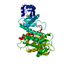
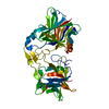
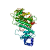
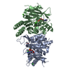
| Entry | Database: PDB / ID: 1avg | ||||||
|---|---|---|---|---|---|---|---|
| Title | THROMBIN INHIBITOR FROM TRIATOMA PALLIDIPENNIS | ||||||
 Components Components |
| ||||||
 Keywords Keywords | COMPLEX (BLOOD COAGULATION/INHIBITOR) / BOVINE THROMBIN / THROMBIN INHIBITOR / COMPLEX (BLOOD COAGULATION-INHIBITOR) / COMPLEX (BLOOD COAGULATION-INHIBITOR) complex | ||||||
| Function / homology |  Function and homology information Function and homology informationsymbiont-mediated perturbation of host defenses /  fibrinogen binding / thrombin / protein polymerization / positive regulation of blood coagulation / acute-phase response / serine-type endopeptidase inhibitor activity / platelet activation / blood coagulation / collagen-containing extracellular matrix ...symbiont-mediated perturbation of host defenses / fibrinogen binding / thrombin / protein polymerization / positive regulation of blood coagulation / acute-phase response / serine-type endopeptidase inhibitor activity / platelet activation / blood coagulation / collagen-containing extracellular matrix / serine-type endopeptidase activity / calcium ion binding / proteolysis / extracellular space / extracellular region fibrinogen binding / thrombin / protein polymerization / positive regulation of blood coagulation / acute-phase response / serine-type endopeptidase inhibitor activity / platelet activation / blood coagulation / collagen-containing extracellular matrix ...symbiont-mediated perturbation of host defenses / fibrinogen binding / thrombin / protein polymerization / positive regulation of blood coagulation / acute-phase response / serine-type endopeptidase inhibitor activity / platelet activation / blood coagulation / collagen-containing extracellular matrix / serine-type endopeptidase activity / calcium ion binding / proteolysis / extracellular space / extracellular regionSimilarity search - Function | ||||||
| Biological species |  Triatoma pallidipennis (insect) Triatoma pallidipennis (insect) Bos taurus (cattle) Bos taurus (cattle) | ||||||
| Method | X-RAY DIFFRACTION / MOLECULAR REPLACEMENT / Resolution: 2.6 Å | ||||||
 Authors Authors | Fuentes-Prior, P. / Huber, R. / Bode, W. | ||||||
 Citation Citation | Journal: Proc.Natl.Acad.Sci.USA / Year: 1997 Title: Structure of the thrombin complex with triabin, a lipocalin-like exosite-binding inhibitor derived from a triatomine bug. Authors: Fuentes-Prior, P. / Noeske-Jungblut, C. / Donner, P. / Schleuning, W.D. / Huber, R. / Bode, W. | ||||||
| History |
|
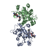
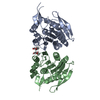
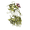
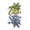
- Structure visualization
| Structure viewer | Molecule: MolmilJmol/JSmol |
|---|
- Downloads & links
-Download
| PDBx/mmCIF format | 1avg.cif.gz | 106 KB | Display | PDBx/mmCIF format |
|---|---|---|---|---|
| PDB format | pdb1avg.ent.gz | 80.9 KB | Display | PDB format |
| PDBx/mmJSON format | 1avg.json.gz | Tree view | PDBx/mmJSON format | |
| Others |  Other downloads Other downloads |
-Validation report
| Arichive directory | https://data.pdbj.org/pub/pdb/validation_reports/av/1avgftp://data.pdbj.org/pub/pdb/validation_reports/av/1avg | HTTPS FTP |
|---|
-Related structure data
| Similar structure data |
|---|

-Links
 PDBj
PDBj


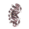
- Assembly
| Deposited unit | 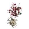
| ||||||||
|---|---|---|---|---|---|---|---|---|---|
| 1 |
| ||||||||
| Unit cell |
|
-Components
| #1: Protein/peptide | Mass: 4792.293 Da / Num. of mol.: 1 / Source method: isolated from a natural source Details: BOVINE THROMBIN WAS PURIFIED FROM FROM FRESH OX BLOOD ACCORDING TO REPORTED PROTOCOLS Source: (natural) Bos taurus (cattle) / Organ: BLOOD / Secretion: BLOOD / Tissue: BLOOD / References: UniProt: P00735, thrombin |
|---|---|
| #2: Protein | Mass: 29772.422 Da / Num. of mol.: 1 / Source method: isolated from a natural source Details: BOVINE THROMBIN WAS PURIFIED FROM FROM FRESH OX BLOOD ACCORDING TO REPORTED PROTOCOLS Source: (natural) Bos taurus (cattle) / Organ: BLOOD / Secretion: BLOOD / Tissue: BLOOD / References: UniProt: P00735, thrombin |
| #3: Protein | Mass: 16039.603 Da / Num. of mol.: 1 Source method: isolated from a genetically manipulated source Source: (gene. exp.) Triatoma pallidipennis (insect) / Organ: BLOOD / Production host: INSECT CELLS / References: UniProt: Q27049 |
| #4: Water | ChemComp-HOH / Water Mass: 18.015 Da / Num. of mol.: 171 / Source method: isolated from a natural source / Formula: H2O Mass: 18.015 Da / Num. of mol.: 171 / Source method: isolated from a natural source / Formula: H2O |
| Compound details | ARG 15 A IS INSERTED IN THE ACTIVE SITE OF A NEIGHBORIN |
-Experimental details
-Experiment
| Experiment | Method: X-RAY DIFFRACTION / Number of used crystals: 2 |
|---|
- Sample preparation
| Crystal | Density Matthews: 2.5 Å3/Da / Density % sol: 50 % | ||||||||||||||||||||||||||||||||||||||||||
|---|---|---|---|---|---|---|---|---|---|---|---|---|---|---|---|---|---|---|---|---|---|---|---|---|---|---|---|---|---|---|---|---|---|---|---|---|---|---|---|---|---|---|---|
| Crystal grow | pH: 4.6 Details: 50 MM NA ACETATE, PH 4.6, 100 MM (NH4)2SO4, 16 % PEG 4,000 | ||||||||||||||||||||||||||||||||||||||||||
| Crystal grow | *PLUS Temperature: 20 ℃ / Method: vapor diffusion, sitting drop | ||||||||||||||||||||||||||||||||||||||||||
| Components of the solutions | *PLUS
|
-Data collection
| Diffraction | Mean temperature: 280 K |
|---|---|
| Diffraction source | Source: ROTATING ANODE / Type: RIGAKU RUH2R / Wavelength: 1.5418 |
| Detector | Type: MARRESEARCH / Detector: IMAGE PLATE / Date: Jun 1, 1996 |
| Radiation | Monochromator: NI FILTER / Monochromatic (M) / Laue (L): M / Scattering type: x-ray |
| Radiation wavelength | Wavelength: 1.5418 Å / Relative weight: 1 |
| Reflection | Resolution: 2.6→19.92 Å / Num. obs: 16392 / % possible obs: 93.2 % / Redundancy: 4.5 % / Rmerge(I) obs: 0.099 / Rsym value: 0.112 / Net I/σ(I): 14.22 |
| Reflection shell | Resolution: 2.6→2.67 Å / Redundancy: 1.9 % / Rmerge(I) obs: 0.121 / Mean I/σ(I) obs: 2.65 / Rsym value: 0.148 / % possible all: 81.6 |
| Reflection | *PLUS Num. measured all: 79890 |
| Reflection shell | *PLUS % possible obs: 81.6 % |
- Processing
| Software |
| ||||||||||||||||||||||||||||||||||||||||||||||||||||||||||||
|---|---|---|---|---|---|---|---|---|---|---|---|---|---|---|---|---|---|---|---|---|---|---|---|---|---|---|---|---|---|---|---|---|---|---|---|---|---|---|---|---|---|---|---|---|---|---|---|---|---|---|---|---|---|---|---|---|---|---|---|---|---|
| Refinement | Method to determine structure: MOLECULAR REPLACEMENT Starting model: BOVINE THROMBIN Resolution: 2.6→10 Å
| ||||||||||||||||||||||||||||||||||||||||||||||||||||||||||||
| Refinement step | Cycle: LAST / Resolution: 2.6→10 Å
| ||||||||||||||||||||||||||||||||||||||||||||||||||||||||||||
| Refine LS restraints |
| ||||||||||||||||||||||||||||||||||||||||||||||||||||||||||||
| Software | *PLUS Name: X-PLOR / Version: 3.842 / Classification: refinement | ||||||||||||||||||||||||||||||||||||||||||||||||||||||||||||
| Refinement | *PLUS | ||||||||||||||||||||||||||||||||||||||||||||||||||||||||||||
| Solvent computation | *PLUS | ||||||||||||||||||||||||||||||||||||||||||||||||||||||||||||
| Displacement parameters | *PLUS | ||||||||||||||||||||||||||||||||||||||||||||||||||||||||||||
| Refine LS restraints | *PLUS
|
