 Movie
Movie Controller

[English] 日本語
 Yorodumi
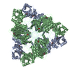
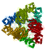
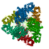
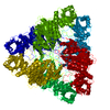
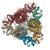
- PDB-1lap: MOLECULAR STRUCTURE OF LEUCINE AMINOPEPTIDASE AT 2.7-ANGSTROMS RE... -
+ Open data

- Basic information
| Entry | Database: PDB / ID: 1lap | ||||||
|---|---|---|---|---|---|---|---|
| Title | MOLECULAR STRUCTURE OF LEUCINE AMINOPEPTIDASE AT 2.7-ANGSTROMS RESOLUTION | ||||||
 Components Components | Cytosol aminopeptidase Leucyl aminopeptidase Leucyl aminopeptidase | ||||||
 Keywords Keywords | HYDROLASE(ALPHA-AMINOACYLPEPTIDE) | ||||||
| Function / homology |  Function and homology information Function and homology informationcysteinylglycine-S-conjugate dipeptidase / prolyl aminopeptidase / leucyl aminopeptidase / dipeptidase activity / metalloaminopeptidase activity / carboxypeptidase activity / disordered domain specific binding / peptidase activity / manganese ion binding / mitochondrion ...cysteinylglycine-S-conjugate dipeptidase / prolyl aminopeptidase / leucyl aminopeptidase / dipeptidase activity / metalloaminopeptidase activity / carboxypeptidase activity / disordered domain specific binding / peptidase activity / manganese ion binding / mitochondrion / proteolysis / cytoplasmSimilarity search - Function | ||||||
| Biological species |  Bos taurus (cattle) Bos taurus (cattle) | ||||||
| Method | X-RAY DIFFRACTION / Resolution: 2.7 Å | ||||||
 Authors Authors | Burley, S.K. / David, P.R. / Taylor, A. / Lipscomb, W.N. | ||||||
 Citation Citation | Journal: Proc.Natl.Acad.Sci.USA / Year: 1990 Title: Molecular structure of leucine aminopeptidase at 2.7-A resolution. Authors: Burley, S.K. / David, P.R. / Taylor, A. / Lipscomb, W.N. #1: Journal: J.Mol.Biol. / Year: 1977Title: Preliminary X-Ray Study of Leucine Aminopeptidase (Bovine Lens), an Oligomeric Metalloenzyme Authors: Jurnak, F. / Rich, A. / Vanloon-Klaassen, L. / Bloemendal, H. / Taylor, A. / Carpenter, F.H. | ||||||
| History |
|
- Structure visualization
| Structure viewer | Molecule: MolmilJmol/JSmol |
|---|
- Downloads & links
-Download
| PDBx/mmCIF format | 1lap.cif.gz | 119.5 KB | Display | PDBx/mmCIF format |
|---|---|---|---|---|
| PDB format | pdb1lap.ent.gz | 93.4 KB | Display | PDB format |
| PDBx/mmJSON format | 1lap.json.gz | Tree view | PDBx/mmJSON format | |
| Others |  Other downloads Other downloads |
-Validation report
| Arichive directory | https://data.pdbj.org/pub/pdb/validation_reports/la/1lapftp://data.pdbj.org/pub/pdb/validation_reports/la/1lap | HTTPS FTP |
|---|
-Related structure data
| Similar structure data |
|---|

-Links
 PDBj
PDBj- Assembly
| Deposited unit | 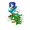
| ||||||||
|---|---|---|---|---|---|---|---|---|---|
| 1 | x 6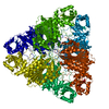
| ||||||||
| Unit cell |
| ||||||||
| Atom site foot note | 1: RESIDUE 471 IS A CIS PROLINE. |
-Components
| #1: Protein | Leucyl aminopeptidase Mass: 52942.098 Da / Num. of mol.: 1 Source method: isolated from a genetically manipulated source Source: (gene. exp.) Bos taurus (cattle) / References: UniProt: P00727, leucyl aminopeptidase | ||
|---|---|---|---|
| #2: Chemical |   Mass: 65.409 Da / Num. of mol.: 2 / Source method: obtained synthetically / Formula: Zn Mass: 65.409 Da / Num. of mol.: 2 / Source method: obtained synthetically / Formula: ZnSequence details | AT THE TIME OF DEPOSITION THE SEQUENCE PRESENTED IN PIR ENTRY APBOL DIFFERS FROM THE SEQUENCE ...AT THE TIME OF DEPOSITION | |
-Experimental details
-Experiment
| Experiment | Method: X-RAY DIFFRACTION |
|---|
- Sample preparation
| Crystal | Density Matthews: 2.91 Å3/Da / Density % sol: 57.76 % |
|---|---|
| Crystal grow | *PLUS Method: vapor diffusion, hanging drop |
| Components of the solutions | *PLUS Conc.: 1-2 M / Chemical formula: Li2SO4 |
-Data collection
| Reflection | *PLUS Highest resolution: 2.7 Å / Lowest resolution: 20 Å / Num. obs: 16334 / Num. measured all: 275554 / Rmerge(I) obs: 0.102 |
|---|
- Processing
| Software | Name: X-PLOR / Classification: refinement | ||||||||||||||||||||||||||||||||||||||||||||||||||||||||||||
|---|---|---|---|---|---|---|---|---|---|---|---|---|---|---|---|---|---|---|---|---|---|---|---|---|---|---|---|---|---|---|---|---|---|---|---|---|---|---|---|---|---|---|---|---|---|---|---|---|---|---|---|---|---|---|---|---|---|---|---|---|---|
| Refinement | Rfactor Rwork: 0.169 / Highest resolution: 2.7 Å | ||||||||||||||||||||||||||||||||||||||||||||||||||||||||||||
| Refinement step | Cycle: LAST / Highest resolution: 2.7 Å
| ||||||||||||||||||||||||||||||||||||||||||||||||||||||||||||
| Refine LS restraints |
| ||||||||||||||||||||||||||||||||||||||||||||||||||||||||||||
| Refinement | *PLUS Highest resolution: 2.7 Å / Rfactor obs: 0.169 | ||||||||||||||||||||||||||||||||||||||||||||||||||||||||||||
| Solvent computation | *PLUS | ||||||||||||||||||||||||||||||||||||||||||||||||||||||||||||
| Displacement parameters | *PLUS | ||||||||||||||||||||||||||||||||||||||||||||||||||||||||||||
| Refine LS restraints | *PLUS Type: x_angle_d |
