 Movie
Movie Controller


+ Open data

- Basic information
| Entry | Database: PDB / ID: 1lan | ||||||
|---|---|---|---|---|---|---|---|
| Title | LEUCINE AMINOPEPTIDASE COMPLEX WITH L-LEUCINAL | ||||||
 Components Components | LEUCINE AMINOPEPTIDASE Leucyl aminopeptidase Leucyl aminopeptidase | ||||||
 Keywords Keywords | HYDROLASE (ALPHA-AMINOACYLPEPTIDE) / AMINOPEPTIDASE / EXOPEPTIDASE / METALLOPEPTIDASE | ||||||
| Function / homology |  Function and homology information Function and homology informationcysteinylglycine-S-conjugate dipeptidase / prolyl aminopeptidase / leucyl aminopeptidase / dipeptidase activity / metalloaminopeptidase activity / carboxypeptidase activity / disordered domain specific binding / peptidase activity / manganese ion binding / mitochondrion ...cysteinylglycine-S-conjugate dipeptidase / prolyl aminopeptidase / leucyl aminopeptidase / dipeptidase activity / metalloaminopeptidase activity / carboxypeptidase activity / disordered domain specific binding / peptidase activity / manganese ion binding / mitochondrion / proteolysis / cytoplasmSimilarity search - Function | ||||||
| Biological species |  Bos taurus (cattle) Bos taurus (cattle) | ||||||
| Method | X-RAY DIFFRACTION / Resolution: 1.9 Å | ||||||
 Authors Authors | Straeter, N. / Lipscomb, W.N. | ||||||
 Citation Citation | Journal: Biochemistry / Year: 1995 Title: Two-metal ion mechanism of bovine lens leucine aminopeptidase: active site solvent structure and binding mode of L-leucinal, a gem-diolate transition state analogue, by X-ray crystallography. Authors: Strater, N. / Lipscomb, W.N. #1: Journal: Biochemistry / Year: 1995Title: Transition State Analogue L-Leucinephosphonic Acid Bound to Bovine Lens Leucine Aminopeptidase: X-Ray Structure at 1.65 Angstroms Resolution in a New Crystal Form Authors: Straeter, N. / Lipscomb, W.N. #2: Journal: Adv.Enzymol.Relat.Areas Mol.Biol. / Year: 1994Title: Structure and Mechanism of Bovine Lens Leucine Aminopeptidase Authors: Kim, H. / Lipscomb, W.N. #3: Journal: J.Mol.Biol. / Year: 1993Title: Re-Refinement of the X-Ray Crystal Structure of Bovine Lens Leucine Aminopeptidase Complexed with Bestatin Authors: Kim, H. / Burley, S.K. / Lipscomb, W.N. #4: Journal: Proc.Natl.Acad.Sci.USA / Year: 1993Title: Differentiation and Identification of the Two Catalytic Metal Binding Sites in Bovine Lens Leucine Aminopeptidase by X-Ray Crystallography Authors: Kim, H. / Lipscomb, W.N. #5: Journal: Biochemistry / Year: 1993Title: X-Ray Crystallographic Determination of the Structure of Bovine Lens Leucine Aminopeptidase Complexed with Amastatin: Formulation of a Catalytic Mechanism Featuring a Gem-Diolate Transition State Authors: Kim, H. / Lipscomb, W.N. #6: Journal: J.Mol.Biol. / Year: 1992Title: Structure Determination and Refinement of Bovine Lens Leucine Aminopeptidase and its Complex with Bestatin Authors: Burley, S.K. / David, P.R. / Sweet, R.M. / Taylor, A. / Lipscomb, W.N. #7: Journal: Proc.Natl.Acad.Sci.USA / Year: 1991Title: Leucine Aminopeptidase: Bestatin Inhibition and a Model for Enzyme-Catalyzed Peptide Hydrolysis Authors: Burley, S.K. / David, P.R. / Lipscomb, W.N. #8: Journal: Proc.Natl.Acad.Sci.USA / Year: 1990Title: Molecular Structure of Leucine Aminopeptidase at 2.7 Angstroms Resolution Authors: Burley, S.K. / David, P.R. / Taylor, A. / Lipscomb, W.N. | ||||||
| History |
|
- Structure visualization
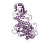
| Structure viewer | Molecule: MolmilJmol/JSmol |
|---|
- Downloads & links
-Download
| PDBx/mmCIF format | 1lan.cif.gz | 114.5 KB | Display | PDBx/mmCIF format |
|---|---|---|---|---|
| PDB format | pdb1lan.ent.gz | 88.2 KB | Display | PDB format |
| PDBx/mmJSON format | 1lan.json.gz | Tree view | PDBx/mmJSON format | |
| Others |  Other downloads Other downloads |
-Validation report
| Arichive directory | https://data.pdbj.org/pub/pdb/validation_reports/la/1lanftp://data.pdbj.org/pub/pdb/validation_reports/la/1lan | HTTPS FTP |
|---|
-Related structure data

-Links
 PDBj
PDBj


- Assembly
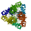
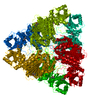
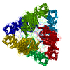
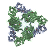
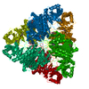
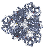
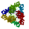
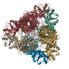
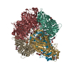
| Deposited unit | 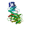
| ||||||||||||||||||
|---|---|---|---|---|---|---|---|---|---|---|---|---|---|---|---|---|---|---|---|
| 1 | x 6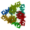
| ||||||||||||||||||
| Unit cell |
| ||||||||||||||||||
| Atom site foot note | 1: CIS PROLINE - PRO A 471 | ||||||||||||||||||
| Components on special symmetry positions |
|
-Components
| #1: Protein | Leucyl aminopeptidase / CYTOSOLIC AMINOPEPTIDASE Mass: 52668.855 Da / Num. of mol.: 1 / Source method: isolated from a natural source / Source: (natural) Bos taurus (cattle) / Organ: LENS / References: UniProt: P00727, leucyl aminopeptidase | ||||||
|---|---|---|---|---|---|---|---|
| #2: Chemical |   Mass: 65.409 Da / Num. of mol.: 3 / Source method: obtained synthetically / Formula: Zn Mass: 65.409 Da / Num. of mol.: 3 / Source method: obtained synthetically / Formula: Zn#3: Chemical | ChemComp-LEU / | Leucine  Type: L-peptide linking / Mass: 131.173 Da / Num. of mol.: 1 / Source method: obtained synthetically / Formula: C6H13NO2 Type: L-peptide linking / Mass: 131.173 Da / Num. of mol.: 1 / Source method: obtained synthetically / Formula: C6H13NO2#4: Chemical | 2-Methyl-2,4-pentanediol  Mass: 118.174 Da / Num. of mol.: 3 / Source method: obtained synthetically / Formula: C6H14O2 / Comment: precipitant*YM Mass: 118.174 Da / Num. of mol.: 3 / Source method: obtained synthetically / Formula: C6H14O2 / Comment: precipitant*YM#5: Water | ChemComp-HOH / | Water Mass: 18.015 Da / Num. of mol.: 384 / Source method: isolated from a natural source / Formula: H2O Mass: 18.015 Da / Num. of mol.: 384 / Source method: isolated from a natural source / Formula: H2O |
-Experimental details
-Experiment
| Experiment | Method: X-RAY DIFFRACTION / Number of used crystals: 1 |
|---|
- Sample preparation
| Crystal | Density Matthews: 2.85 Å3/Da / Density % sol: 56.85 % | ||||||||||||||||||||||||||||||||||||||||||
|---|---|---|---|---|---|---|---|---|---|---|---|---|---|---|---|---|---|---|---|---|---|---|---|---|---|---|---|---|---|---|---|---|---|---|---|---|---|---|---|---|---|---|---|
| Crystal grow | pH: 7.8 / Details: pH 7.8 | ||||||||||||||||||||||||||||||||||||||||||
| Crystal grow | *PLUS Method: vapor diffusion, hanging drop | ||||||||||||||||||||||||||||||||||||||||||
| Components of the solutions | *PLUS
|
-Data collection
| Diffraction | Mean temperature: 123 K |
|---|---|
| Diffraction source | Wavelength: 1.5418 Å |
| Detector | Type: XENTRONICS / Detector: AREA DETECTOR / Date: Jun 6, 1995 |
| Radiation | Monochromator: SUPPER DOUBLE MIRRORS / Monochromatic (M) / Laue (L): M / Scattering type: x-ray |
| Radiation wavelength | Wavelength: 1.5418 Å / Relative weight: 1 |
| Reflection | Num. obs: 44837 / % possible obs: 99.3 % / Redundancy: 3.6 % / Rmerge(I) obs: 0.07 |
| Reflection | *PLUS Highest resolution: 1.9 Å / Num. measured all: 160257 / Rmerge(I) obs: 0.07 |
- Processing
| Software |
| ||||||||||||||||||||||||||||||||||||||||||||||||||||||||||||
|---|---|---|---|---|---|---|---|---|---|---|---|---|---|---|---|---|---|---|---|---|---|---|---|---|---|---|---|---|---|---|---|---|---|---|---|---|---|---|---|---|---|---|---|---|---|---|---|---|---|---|---|---|---|---|---|---|---|---|---|---|---|
| Refinement | Resolution: 1.9→7 Å / σ(F): 2
| ||||||||||||||||||||||||||||||||||||||||||||||||||||||||||||
| Displacement parameters | Biso mean: 16.3 Å2 | ||||||||||||||||||||||||||||||||||||||||||||||||||||||||||||
| Refine analyze | Luzzati coordinate error obs: 0.2 Å | ||||||||||||||||||||||||||||||||||||||||||||||||||||||||||||
| Refinement step | Cycle: LAST / Resolution: 1.9→7 Å
| ||||||||||||||||||||||||||||||||||||||||||||||||||||||||||||
| Refine LS restraints |
| ||||||||||||||||||||||||||||||||||||||||||||||||||||||||||||
| Software | *PLUS Name: X-PLOR / Version: 3.1 / Classification: refinement | ||||||||||||||||||||||||||||||||||||||||||||||||||||||||||||
| Refine LS restraints | *PLUS
| ||||||||||||||||||||||||||||||||||||||||||||||||||||||||||||
| LS refinement shell | *PLUS Highest resolution: 1.9 Å / Lowest resolution: 2 Å |
