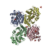
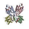
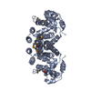
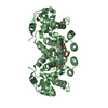
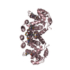
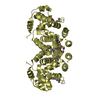
A: Putative ubiquinone biosynthesis protein B: Putative ubiquinone biosynthesis protein C: Putative ubiquinone biosynthesis protein D: Putative ubiquinone biosynthesis protein
Mass: 25790.289 Da / Num. of mol.: 4 Source method: isolated from a genetically manipulated source Source: (gene. exp.) Nostoc punctiforme (bacteria) / Strain: ATCC 29133 / PCC 73102 / Gene: 23130561, Npun_R0350 / Plasmid: SpeedET / Production host: Escherichia Coli (E. coli) / Strain (production host): HK100 / References: UniProt: B2J5W7
Sequence details
THE CONSTRUCT WAS EXPRESSED WITH A PURIFICATION TAG MGSDKIHHHHHHENLYFQG. THE TAG WAS REMOVED WITH ...THE CONSTRUCT WAS EXPRESSED WITH A PURIFICATION TAG MGSDKIHHHHHHENLYFQG. THE TAG WAS REMOVED WITH TEV PROTEASE LEAVING ONLY A GLYCINE (0) FOLLOWED BY THE TARGET SEQUENCE.
-
Experimental details
-
Experiment
Experiment
Method: X-RAY DIFFRACTION / Number of used crystals: 1
-
Sample preparation
Crystal
Density Matthews: 3.05 Å3/Da / Density % sol: 59.65 % Description: DATA WERE SCALED USING XSCALE WITH FRIEDEL PAIRS KEPT AS SEPARATE WHEN COMPUTING R-SYM, COMPLETENESS AND
Crystal grow
Temperature: 293 K / Method: vapor diffusion, sitting drop / pH: 5.5 Details: 20.0000% polyethylene glycol 3000, 0.1M citric acid pH 5.5, Additive: 0.001 M Ubiquinone, NANODROP', VAPOR DIFFUSION, SITTING DROP, temperature 293K
Type: MARMOSAIC 325 mm CCD / Detector: CCD / Date: Jun 15, 2008 / Details: Flat mirror (vertical focusing)
Radiation
Monochromator: Single crystal Si(111) bent monochromator (horizontal focusing) Protocol: MAD / Monochromatic (M) / Laue (L): M / Scattering type: x-ray
Radiation wavelength
ID
Wavelength (Å)
Relative weight
1
0.91837
1
2
0.97937
1
3
0.97882
1
Reflection
Resolution: 2.85→49.266 Å / Num. obs: 26383 / % possible obs: 82.4 % / Observed criterion σ(I): -3 / Biso Wilson estimate: 86.669 Å2 / Rmerge(I) obs: 0.093 / Net I/σ(I): 9.23
Reflection shell
Resolution (Å)
Rmerge(I) obs
Mean I/σ(I) obs
Num. measured obs
Num. unique obs
% possible all
2.85-2.92
1.11
0.99
8232
3567
83.8
2.92-3
0.839
1.28
7846
3407
83.8
3-3.09
0.688
1.61
7652
3311
83.4
3.09-3.19
0.463
2.31
7499
3250
83.5
3.19-3.29
0.332
3.15
7403
3163
83.4
3.29-3.41
0.261
4.12
7076
3032
82.7
3.41-3.54
0.189
5.38
6667
2878
83.1
3.54-3.68
0.132
7.51
6571
2793
82.9
3.68-3.84
0.105
9.36
6221
2652
82.4
3.84-4.03
0.085
11.1
6022
2560
82.2
4.03-4.25
0.069
13.4
5714
2415
81.8
4.25-4.51
0.055
15.3
5337
2258
81.6
4.51-4.82
0.048
17.3
5080
2139
81.3
4.82-5.2
0.049
16.8
4706
1975
81.7
5.2-5.7
0.052
17.4
4327
1823
81.3
5.7-6.37
0.048
17.3
3948
1651
81.2
6.37-7.36
0.041
20.2
3468
1451
81.3
7.36-9.01
0.035
24.8
2916
1207
80.3
9.01-12.75
0.027
30.9
2233
931
80.4
12.75-49.27
0.028
31
1152
488
75.4
-
Phasing
Phasing
Method: MAD
-
Processing
Software
Name
Version
Classification
NB
MolProbity
3beta29
modelbuilding
PHENIX
refinement
SHELX
phasing
XSCALE
datascaling
PDB_EXTRACT
3.006
dataextraction
XDS
datareduction
SHELXD
phasing
autoSHARP
phasing
BUSTER
2.8.0
refinement
Refinement
Method to determine structure: MAD / Resolution: 2.85→49.266 Å / Cor.coef. Fo:Fc: 0.937 / Cor.coef. Fo:Fc free: 0.912 / Occupancy max: 1 / Occupancy min: 0.75 / Cross valid method: THROUGHOUT / σ(F): 0 Details: 1. A MET-INHIBITION PROTOCOL WAS USED FOR SELENOMETHIONINE INCORPORATION DURING PROTEIN EXPRESSION. THE OCCUPANCY OF THE SE ATOMS IN THE MSE RESIDUES WAS REDUCED TO 0.75 FOR THE REDUCED ...Details: 1. A MET-INHIBITION PROTOCOL WAS USED FOR SELENOMETHIONINE INCORPORATION DURING PROTEIN EXPRESSION. THE OCCUPANCY OF THE SE ATOMS IN THE MSE RESIDUES WAS REDUCED TO 0.75 FOR THE REDUCED SCATTERING POWER DUE TO PARTIAL S-MET INCORPORATION. 2. B-FACTORS CONTAIN BOTH TLS AND RESIDUAL COMPONENTS.
In the structure databanks used in Yorodumi, some data are registered as the other names, "COVID-19 virus" and "2019-nCoV". Here are the details of the virus and the list of structure data.
Jan 31, 2019. EMDB accession codes are about to change! (news from PDBe EMDB page)
EMDB accession codes are about to change! (news from PDBe EMDB page)
The allocation of 4 digits for EMDB accession codes will soon come to an end. Whilst these codes will remain in use, new EMDB accession codes will include an additional digit and will expand incrementally as the available range of codes is exhausted. The current 4-digit format prefixed with “EMD-” (i.e. EMD-XXXX) will advance to a 5-digit format (i.e. EMD-XXXXX), and so on. It is currently estimated that the 4-digit codes will be depleted around Spring 2019, at which point the 5-digit format will come into force.
The EM Navigator/Yorodumi systems omit the EMD- prefix.
Related info.:Q: What is EMD? / ID/Accession-code notation in Yorodumi/EM Navigator
Yorodumi is a browser for structure data from EMDB, PDB, SASBDB, etc.
This page is also the successor to EM Navigator detail page, and also detail information page/front-end page for Omokage search.
The word "yorodu" (or yorozu) is an old Japanese word meaning "ten thousand". "mi" (miru) is to see.
Related info.:EMDB / PDB / SASBDB / Comparison of 3 databanks / Yorodumi Search / Aug 31, 2016. New EM Navigator & Yorodumi / Yorodumi Papers / Jmol/JSmol / Function and homology information / Changes in new EM Navigator and Yorodumi
 Movie
Movie Controller

 Yorodumi
 Open data
 Basic information
 Components
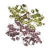
 Coenzyme Q10
  Keywords
 Function and homology information
 Authors
 Citation
 Structure visualization
 Downloads & links
 Other downloads
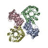


 PDBj
PDBj Assembly


 Sample preparation
 / Beamline: BL11-1 / Wavelength: 0.91837,0.97937,0.97882
 Processing